Biological Classification
2.1 Systems of Classification
2.2 Classification of Organisms
2.3 Kingdom Monera
2.4 Kingdom Protista
2.5 Kingdom Mycota
2.6 Kingdom Plantae
2.7 Kingdom Animalia
2.8 Prions, Viroids, Viruses and Lichens
Biological classification has an ancient history and is closely associated with human evolution. Classification is a human invention to create order in the tremendous biodiversity of life forms. Over the period of time we have discovered a million and a half different types of organisms and many more arc to be discovered from rain forests, oceans and other parts of the earth. Human beings classified or grouped plants into harmful and useful ones, non-edible and edible, medicinally useful, source of timber and fibre yielding plants.
The process of classification is a constantly continuing process because there are many plants and animals which are taxonomic puzzles.
2.1 SYSTEMS OF CLASSIFICATION
The different and numerous systems of classification developed can be moped into artificial, natural and phylogenetic.
2.1.1 Artificial systems
This system is the oldest one and it uses one or very few characters to lassify. There is no consideration of evolutionary relationships.
Aristotle in 350 B.C. classified all things on earth into plants, Animais and minerals. He also classified organisms on the basis of where they live, into aquatic (water) and terrestrial (land). Theophrastus who was a student of Aristotle classified plants into herbs, shrubs and trees based upon their appearance or habit. Charaka (1501 B.C.) classified medicinal plants.
In 1753 Linnaeus, the father of taxonomy classified plants by a sexual character of number of stamens in the flower into mondria, diandria, triandria and so on. It was a artificial system since only one character was considered.
Artificial systems are easy to create but have the following demerits.
1 They do not indicate any evolutionary relationships.
2 Different types of organisms like bird, bat, insects are grouped together.
3 Organisms which have common characters get separated like whale, horse and bat.
Linnaeus classified organisms into two kingdoms namely Plantae (Plants) and Animalia I (Animals)
2.1.2 Natural systems
In this system many characters are taken into consideration for classifying organisms. In classifying I plants various factors like chromosomal morphology, anatomy of parts, embryology, biochemistry are I considered and maximum number of similarities and differences are considered for classification. Fori example mammals have a set of similar characters like mammary glands, body hair, a heart of four! chambers and are warm blooded.
In plants the type of flower, all aspects of nature of petals, stamens, carpels, presence or absence I of floral disc, position of ovary in the flowers superior, inferior or semi-inferior, were all considered I When Bentham and Hooker (Fig. 2.2 and 2.3) classified flowering plants all the above mentioned and! many more characters were considered.
George Bentham and Dalton Hooker published a three-volume work titled Genera Plantarum. I The natural systems are pre-Darwinian, therefore there was no study of evolutionary relationships. (Post Darwinian systems took into consideration Darwinian concepts of organic evolution). Some important features of natural systems are-
1 Many morphological characters are considered.
2 Many homologies of characters like morphology (external structure), anatomy (internal structure), cytotaxonomy, biochemistry are considered. Homology is the study of the relationship of comparable structures including biomolecules like proteins and nucleic acids.
3 Related organisms are grouped together.
4 The system enables one to identify plants very quickly because the sets of characters of different taxa are well-delineated. The system provides efficient artificial keys for identification.
2.1.3 Phylogenetic systems
These systems are based on evolutionary process. Apart from classifying plants the system traces out the evolutionary relations, organisms of the same group are considered to have evolved from a common ancestor. Darwin’s theory of organic evolution has tremendously influenced these systems. The phylogenetic classification of angiosperms by all taxonomists lay more emphasis on floral characters since these characters are highly conserved and therefore they are reliable constants for classification.
Some importance is also given to unique vegetative characters seen in plants of some taxonomic groups.
One of the first phylogenetic systems proposed was by two German botanists Engler and Prantl (1887 - 99) in their magnum opus publication in German ‘Die Naturalichen Pflanzen Familien’. Their system involved all plants from algae to angiosperms.
The merits of the system are that it is an exhaustive work, the classification is based on evolutionary concepts, all different characters like morphology, anatomy, cytology and embryology were considered and they placed gymnosperms before angiosperms. (Bentham and Hooker had wronglyplaced gymnosperms between monocots and dicots). Another merit is that it is a progressive system should have been placed after dicots. They considered unisexual flowers as primitive and bisexual ones as advanced. This is not true since we have evidences to prove that unisexual flowers have originated from bisexual ones. The presence of sterile stamens (staminodes) in unisexual female flowers and presence of sterile pistils (pistillodes) in unisexual male flowers proves the derived nature or more evolved nature of unisexual flowers. Another demerit is they suggested that all the groups of angiosperms have evolved from one ancestral stock (monophyletic origin), but we have many evidences that angiosperms have evolved from different ancestral stocks (polyphyletic origin).
Hutchinson (1959), Takhtajan (1966) proposed improved phylogenetic systems. A major source of information for phyletic or phylogenetic systems are the fossil records and as more and more fossil angiosperms are discovered by the paleobotanists our knowledge of evolution and phytogeny will improve.
Recent trends in taxonomy (classification) is referred to as phenetic classification. In phenetics organisms are grouped together on the basis of the sum total of their similarities.
In 1940, Julian Huxley introduced the concept termed as new systematics, this concept incorporated old concepts of morphology and added new branches like anatomy, chromosome study, ecology, biochemistry, physiology, genetics of entire populations were employed to create new systematics (modem taxonomy) which is called biosystematics. Earlier systematics or taxonomy where external appearance (morphology) was the only criteria for classification is now called classical systematics.
2.1.4 New systematics
New systematics (modern taxonomy) is a combination of many new branches like numerical taxonomy, cytotaxonomy, chemotaxonomy and cladistics.
Numerical taxonomy : It is also called Adansonian taxonomy after its developer. In this methodology all characters are observed and recorded and numbers are assigned to each character of the plant. These code numbers are later used for computer analysis.
If a character coded for is present in the plant then it is a + sign- If the character coded for is
then it is - sign. If no data on characters is.Available then a Design assigned. Many characters are involved with equal weightage. By computer programming a huge and easily accessible data base of innumerable plants are created. By this method we establish a numerical degree of relationships between plants which will be of great help in taxonomic classification.
Cytotaxonomy : This branch mainly deals with study of chromosomes, their structural differences, behaviour at meiosis, nature of aberrations, if any, are considered while classifying plants. Chromosomal studies have proved ape like ancestors for human evolution. Same chromosome number is seen in different species of the same genus, for example, twelve species of the genus Solatium have identical chromosome numbers.
Chemotaxonomy : The basis for chemotaxonomy is the presence of unique chemicals in plants. Very often when two plants have same unique chemicals they how numerous similarites in other charcters and such parts can be classified or grouped together in common genus, family or order.
These unique chemicals very often are secondary metabolites like betacyanins or even aromatic compounds, for example, if a plant part has sulfurous odour in its sap it belongs to mustard plant family like Brassicaceae which includes related plants like cabbage, turnip and cauliflower. The nucleotide sequences of DNA also establish evolutionary affinities between organisms and the data obtained is of use in phylogenetically classifying organisms. DNA nucleotide sequencing studies have shown that humans are more closely related to chimpanzees than gorillas.
Cladistcs : In this technique we create a phylogenetic tree or a family tree or a cladogram which is a diagrammatic representation of the evolutionary ancestry of different groups of plants or animals (Fig. 2.6). A family tree created on the basis Of numerical taxonomycalled dendrogram. The ancestors of modem groups have ancestral characters and modem groups have developed, modified or derived characters during evolution. Due to the presence of numerous derived characters we have numerous subgroups, for example, a family of plants can have many subfamilies.
In 1967. Armen Takhtajan opined that phylogeny is to taxonomy like what bones are to muscles or flesh.
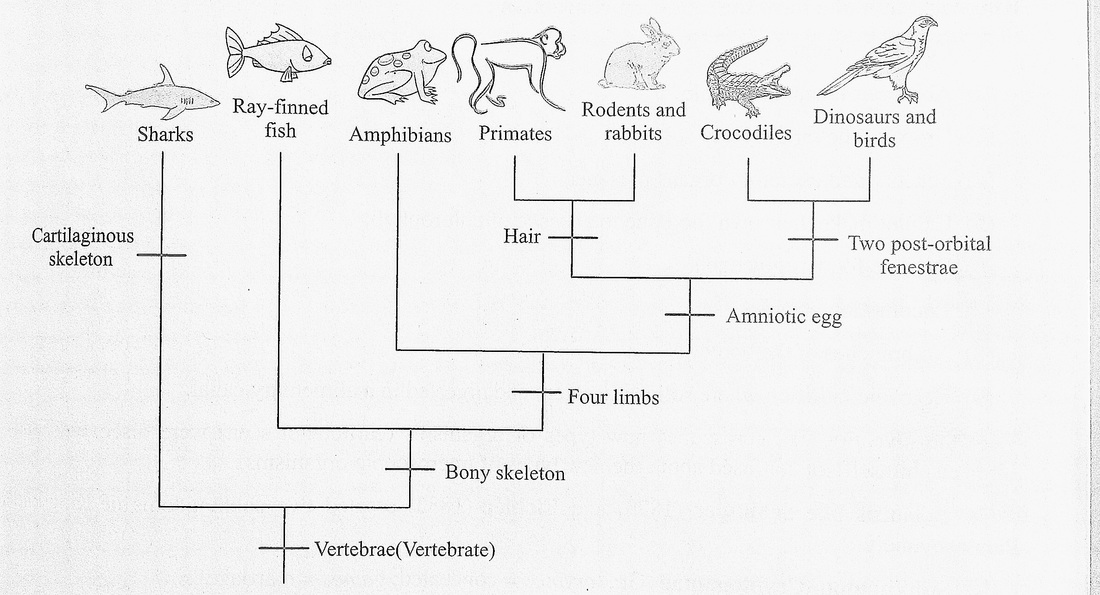
Fig. 2.1 A simple cladogram
2.1 CLASSIFICATION OF ORGANISMS
2.2.1 Two kingdom system
All living forms were classified into two kingdoms namely Kingdom Plantae including all plants and Kingdom Animalia which included all animals. Carolus Linnaeus proposed this classification in 1758.
Kingdom Plantae have the following characters :
4. Cell wall present
5. Central vacuole in the cell.
6. Growth limited to growing points.
7. Absence of sensory and excretory organs.
8. Ability to make food by photosynthesis (presence of chlorophyll).
9. Reserve food as starch.
10. Absence of locomotion.
11. Presence of branches without uniform shape.
12. Water and nutrients taken by absorption.
Kingdom Animalia have the following characters :
(1) Cell wall absent
(2) Absence of central vacuole.
(3) Growth all over the body.
(4) Sensory and excretory organs present
(5) Cannot make their own food due to absence of chlorophyll.
(6) Reserve food as glycogen.
(7) Locomotion occurs.
(8) No branches of the body.
(9) Holozoic nutrition where solids are eaten and digested in a alimentary canal.
With the invention of microscopes new types of organisms (earlier not seen) were discovered and Linnaeus himself got confused about the new types of microscopic organisms.
Botanists like Schimper (1879) and Eichler (1883) created two subkingdoms of Kingdom Plantae namely.
(1) Subkingdom Cryptogamae (Gr., cryptos = concealed; games = marriage)
1 The cryptogams are thallophyta (Gr., thallose = undifferentiated; phyta = plant) like algae and fungi. The thallophyta has a plant body called thallus; it is a simple plant body without well organized tissues. The Bryophyta and Pteridophyta were more evolved than thallophyta since they have well formed tissue systems. Subkingdom Phanerogamae (Gr., phanero = visible; gamos =, marriage or sexual parts)
Phanerogams are the seed bearing plants. Spermatophyta (Gr., sperma = seed; phyta = plant) and the division spermatophyta was made up of two subdivisions namely gymnosperms (Gr., gymno = naked; sperma = seed) and angiosperms (Gr., angio = box; sperma = seed)
In gymnosperms seeds formed from ovules are naked since ovaries are absent In angiosperms the seeds are enclosed in fruit which is produced by the ovary.
Limitations of two kingdom system of Linnaeus
The Linnaean two kingdom system was in acceptance for a long time. With the invention of better and better microscopes a new group of microscopic organisms were discovered and quite a few of them were taxonomic puzzles.
The drawbacks or limitation of the two kingdom could be described as follows.
1. Some microscopic forms like Euglena have no cell wall like animals, but has chlorophyll like plants and it shows holozoic nutrition like animals.
Animals like sponges are fixed without locomotion and are branched regularly like plants and they also have animal characters.
2. The fungi have cell walls like plants but are heterotrophic like animals since they have no chlorophyll. Eventhough they have cell wall it is made up of chitin whereas green plants have a cell wall of cellulose.
3. Organisms like lichens have two components, the fungal component that encloses the algae cells. They could not fit into either of two kingdoms of Linnaeus.
4. Prokaryotes (Gr., pro = before; karyon = kernel, nucleus) were discovered, they do not have nucleus in their cells, the DNA of the chromosomes are not associated with histone proteins (naked DNA), membrane bound organelles like lysosomes, true vacuoles, mitochondria, Golgi bodies are absent. There is no mitosis and meiosis, no spindle fibre formation and no cytoskeletal structures.
In contrast to prokaryotes there are the eukaryotes (Gr., eu = good; karyon = kernel or nucleus' which have nucleus with nuclear membrane and nucleoli, DNA of chromosomes are associated with proteins. All membrane bound organelles absent in prokaryotes occur in eukaryotes. Mitosis and meiosis occur, spindle fibres and cytoskeletal structures are present. It was therefore necessar to separate prokaryotes and eukaryotes.
5. With the invention of electron microscope a whole new world of ultramicroscopic infection particles were discovered, like the viruses and viroids.
Supporters of Linnaeus's two kingdom classification placed prokaryotes and viruses in Kingdom Plantae and viruses were a part of botany syllabus for a long time.
2.2.2 Three Kingdom system
In 1866 Ernst Haeckel created a third kingdom for unicellular animals, algae and fungi. In all the there was no tissue formation. The third kingdom was called Protista by Haeckel. Subsequently fun was removed from this kingdom and Protista included only unicellular microscopic organisms.
2.2.3 Four Kingdom system
With the invention of electron microscope very fine details of bacterial cells Structure (ultrastructure) Was ; Observed These details would not be seen with compound microscopes which had low magnification. |, was observed that bacteria are prokaryotes unlike eukaryotes which have nucleus and other membrane bound organelles. In 1956, Copeland created a new Kingdom Monera for organisms like bacteria and cyanobacteria (blue green algae) all of which were prokaryotic. The fungi were included in Plantae.
2.2.3 Five Kingdom system
In 1969, R.H. Whittaker published an article in scientific journal called ‘Science’ titled ‘New concepts of kingdoms of organisms’ and here he classified all organisms into a five kingdom classification namely Kingdom Monera, Protista, Mycota (fungi), Plantae (Metaphyta), Animalia (Metazoa),
Viruses, viroids and prions have not been assigned to any of these five kingdoms since they have highly unique characters or some uniquely absent characters which will be dealt with in later chapters.
It is now considered that during evolution monerans gave rise to protists and from the protista life forms the mycotans, plantae and animalia forms of life evolved. The mycotans (fungi) first evolved from protists and later on about one billion years in the past some of the protists evolved into animal life forms and around 400 million years ago from photosynthetic protists the plantae lifeforms evolved (Fig. 2.7).
2.2.4.1 Salient features of the five kingdoms Kingdom Monera (The prokaryotes)
The kingdom includes prokaryotes called monerans (bacteria, cyanobacteria). They have following salient features or characteristics.
1 Simplest or most primitive organisms which are prokaryotic (without nucleus) and DNA is naked.
2 The cell walls have peptidoglycan or murein (no cellulose).
3 The membrane bound cell organelles are absent. .
4 Ribosomes are of 50S and 30S subunits. During protein synthesis they associate to form 70S complexes.(S = Svedberg unit which is sedimentation coefficient)
5 They have all types of nutritional patterns like saprophytic, parasitic, chemoautotrophic or chemosynthetic, photoautotrophic or photosynthetic and symbiotic.
6 Flagellum is one stranded, unlike the 11 stranded flagellum of eukaryotes.
7 Reproduction by vegetative, asexual and parasexual methods (the partial transfer of genetic material).
8 Some have the ability to fix nitrogen into useful nitrates (nitrogen fixation).
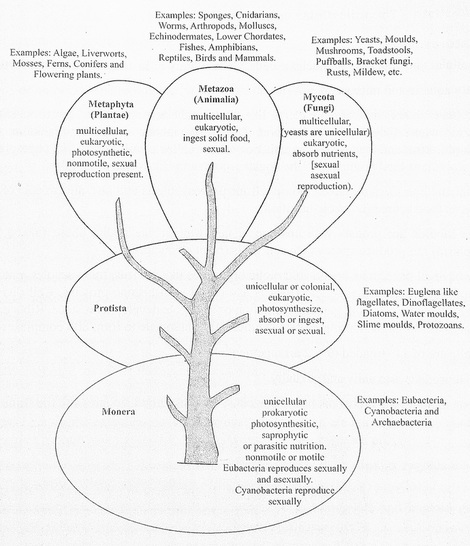
Fig. 2.2 The five Kingdoms and the probable lines of evolution
Kingdom Protista (The unicellular eukaryotes)
Salient features
1 Unicellular and generally uninucleate cells. Cells solitary or in colonies (no tissue formation).
2 Mostly aquatic and microscopic.
3 They have diverse nutritional patterns like photosynthesis, saprophytic, parasitic, ingestive or holozoic where they take in solid food. The photosynthetic unicellular planktons are called phytoplankton and the non-photosynthetic ones are called zooplankton. The phytoplankton are very important food producers of the aquatic ecosystems like oceans and lakes.
Organisms like Euglena have chlorophyll for photosynthesis and also are capable of ingesting solids as food source (holozoic nutrition).
4 Slime moulds are protists with absence of cell wall in plasmodial phase (naked amoeboid cytoplasm) but produce spores with cell walls (plant character).
5 They have all membrane bound eukaryotic organelles like nucleus, true vacuoles, mitochondria, endoplasmic reticulum etc., The DNA is associated with histone proteins. Cell division is by mitosis. Meiosis occurs at gametic formation or at zygote develoument stage. Ribosomes are of 60S and 40S subunits. During protein synthesis they associate to form 80S complexes.
6 Flagella are of 11 stranded (9 + 2 arrangement).
7 They reproduce asexually and sexually.
Protista includes the protistans like protozoans, diatoms, dinoflagellates and the slime moulds.

Kingdom Mycota (generally multicellular decomposers)
This kingdom includes the mycotans represented by the fungi.
Salient features
1. 1 Plant body is made up of cylindrical thread like structures called hyphae (sing., hypha). Numerous hyphae coil around one another to form the mycelia (sing., mycelium). The hyphae could be aseptate or septate and the cells binucleate or multinucleate. Yeasts are unicellular.
2 The cell wall is made up of chitin.
3 Cell structure typically eukaryotic as in Protista.
4 Reserve food is glycogen.
5 Mode of nutrition is heterotrophic (saprophytic or parasitic).
6 Reproduction is by asexual and sexual methods. Asexual by motile spores (zoospores) or by non-motile spores (aplanospores) or conidia. In lower fungi like Phycomycetes sex organs like the unicellular antheridia (male) and unicellular oogonia(female) are produced. In higher fungi like Basidiomycetes sexual process is present but sex organs are absent. Ascomycete and Basidiomycete fungi produce fruiting bodies by sexual process called ascocarps and basidiocarps respectively during sexual process.
Some common fungi are moulds, mildews, smuts, rusts, bracket fungi, morels, mushrooms and yeasts which are the unicellular sugar fungi.
Kingdom Plantae or Metaphyta (Autotrophs or producers of organic food)
They include higher algae, bryophytes, pteridophytes and spermatophytes (gymnosperms and angiosperms).
Salient features
1 Mostly anchored to soil by rhizoids or roots, some are aquatic.
2 Cellulose present in cell wall, tissue and organ level of organisation present
3 Autotrophic or photosynthetic with a few exceptions which are parasitic. Some autotrophs like Nepenthes capture and digests insects (carnivorous or insectivorous plants).
4 Reproduction is asexual and sexual. Except for algae, all have diploid zygotes that divide by mitosis to form diploid embryos.
5 There is alternation of generations between a diploid sporophyte and haploid gametophyte generation (except most algae).
6 Sporophytes of pteridophytes and spermatophytes have developed vascular tissues like xylem and phloem for conduction.
Kingdom Animalia or Metazoa (Hetrotrophs or consumers of organic food)
This kingdom is of multicellular consumers or phagotrophs who eat solid food (holozoic) which is digested in the alimentary canal.
1 All prokaryotes have been assigned to a separate Kingdom Monera.
2 All unicellular eukaryotes in Kingdom Protista.
3 A separate kingdom Mycota for fungi.
4 Plantae and Animalia clearly delineated.
5 The classification has attempted to study the evolutionary or phyletic relationships between members of the different kingdoms.
2.2.4.3Demerits or drawbacks of the five kingdom classification
2.2 KINGDOM - MONERA
In this chapter we will study in some detail monerans like Bacteria, Cyanobacteria, Rickettsias, Actinomycetes and Mycoplasma.
2.3.1 Bacteria
It was Anton Leeuwenhoek who observed bacteria from pond water and tartar of teeth in 1674. He saw them using microscopes made by him.The term bacteria was proposed by Ehrenberg in 1829.
Distribution
They are ubiquitous/cosmopolitan in distribution. They are found in air, soil and water. The reasons why I they are found in all habitats is their small size, rapid reproductive rate, numerous modes of nutrition and formation of air-borne endospores. Bacterial cells have been seen in sea beds at four kilometre depth and in atmosphere at a height of six kilometres.
Size
These microscopic organisms have a cell size with a length of 1.5 um and width of 0.5 um in rod shaped cells. The spherical cells have a diameter of 1um to 5um. Spiral shaped cells are the largest sized reaching upto 20um or more in length.
Classification of bacteria based on cell shape or morphology
1. Cocci (sing., coccus): They are oval or spherical cells without flagella. The spheres occur as single cells (monococcus), a pair of cells (diplococcus), in groups of four cells or tetrads (tetracoccus) or as a chain of cells (streptococcus), in clusters (staphylococcus) or cells in a cube like arrangement called sarcina.
2. Bacilli (sing., bacillus): They are rod shaped cells which may occur singly (monobacillus), in pairs (diplobacillus), in chains (streptobacillus), or as a layer with many cells called palisade bacillus.
This kingdom includes the mycotans represented by the fungi.
Salient features
1. 1 Plant body is made up of cylindrical thread like structures called hyphae (sing., hypha). Numerous hyphae coil around one another to form the mycelia (sing., mycelium). The hyphae could be aseptate or septate and the cells binucleate or multinucleate. Yeasts are unicellular.
2 The cell wall is made up of chitin.
3 Cell structure typically eukaryotic as in Protista.
4 Reserve food is glycogen.
5 Mode of nutrition is heterotrophic (saprophytic or parasitic).
6 Reproduction is by asexual and sexual methods. Asexual by motile spores (zoospores) or by non-motile spores (aplanospores) or conidia. In lower fungi like Phycomycetes sex organs like the unicellular antheridia (male) and unicellular oogonia(female) are produced. In higher fungi like Basidiomycetes sexual process is present but sex organs are absent. Ascomycete and Basidiomycete fungi produce fruiting bodies by sexual process called ascocarps and basidiocarps respectively during sexual process.
Some common fungi are moulds, mildews, smuts, rusts, bracket fungi, morels, mushrooms and yeasts which are the unicellular sugar fungi.
Kingdom Plantae or Metaphyta (Autotrophs or producers of organic food)
They include higher algae, bryophytes, pteridophytes and spermatophytes (gymnosperms and angiosperms).
Salient features
1 Mostly anchored to soil by rhizoids or roots, some are aquatic.
2 Cellulose present in cell wall, tissue and organ level of organisation present
3 Autotrophic or photosynthetic with a few exceptions which are parasitic. Some autotrophs like Nepenthes capture and digests insects (carnivorous or insectivorous plants).
4 Reproduction is asexual and sexual. Except for algae, all have diploid zygotes that divide by mitosis to form diploid embryos.
5 There is alternation of generations between a diploid sporophyte and haploid gametophyte generation (except most algae).
6 Sporophytes of pteridophytes and spermatophytes have developed vascular tissues like xylem and phloem for conduction.
Kingdom Animalia or Metazoa (Hetrotrophs or consumers of organic food)
This kingdom is of multicellular consumers or phagotrophs who eat solid food (holozoic) which is digested in the alimentary canal.
- Multicellular tissue forming eukaryotes with different organs and systems like digestive, nervous, excretory, respiratory and reproductive systems.
- Cell wall absent.
- Mechanical support by means of a well-developed skeletal system, muscles and ligaments.
- Reproduction is sexual with embryo formation.
- It is a large kingdom divided into numerous phyla (Details of which you will study in chapter 4).
1 All prokaryotes have been assigned to a separate Kingdom Monera.
2 All unicellular eukaryotes in Kingdom Protista.
3 A separate kingdom Mycota for fungi.
4 Plantae and Animalia clearly delineated.
5 The classification has attempted to study the evolutionary or phyletic relationships between members of the different kingdoms.
2.2.4.3Demerits or drawbacks of the five kingdom classification
- Even in a kingdom there are highly diverse groups, like in monerans, we have organisms with and without cell wall, organisms that are unicellular and with colonies like filament of cells.
- Archaebacteria were included in Monera but now they are in separate domain, since these ancient Bacteria are different from Bacteria of Monera.
- Mycoplasma have unique features but they are in the Kingdom Monera.
- There is no place for viruses, viroids and prions in the five kingdom classification.
- Currently we have the three domains of the living world namely domain Archaea with archaebacteria, domain Bacteria (Gr.,pro = before; karyon = kernel or nucleus ) with eubacteria. cyanobacteria and the domain Eukarya (Gr., eu = true; karyon = kernel or nucleus) with Protista, Plantae and Animalia.
2.2 KINGDOM - MONERA
In this chapter we will study in some detail monerans like Bacteria, Cyanobacteria, Rickettsias, Actinomycetes and Mycoplasma.
2.3.1 Bacteria
It was Anton Leeuwenhoek who observed bacteria from pond water and tartar of teeth in 1674. He saw them using microscopes made by him.The term bacteria was proposed by Ehrenberg in 1829.
Distribution
They are ubiquitous/cosmopolitan in distribution. They are found in air, soil and water. The reasons why I they are found in all habitats is their small size, rapid reproductive rate, numerous modes of nutrition and formation of air-borne endospores. Bacterial cells have been seen in sea beds at four kilometre depth and in atmosphere at a height of six kilometres.
Size
These microscopic organisms have a cell size with a length of 1.5 um and width of 0.5 um in rod shaped cells. The spherical cells have a diameter of 1um to 5um. Spiral shaped cells are the largest sized reaching upto 20um or more in length.
Classification of bacteria based on cell shape or morphology
1. Cocci (sing., coccus): They are oval or spherical cells without flagella. The spheres occur as single cells (monococcus), a pair of cells (diplococcus), in groups of four cells or tetrads (tetracoccus) or as a chain of cells (streptococcus), in clusters (staphylococcus) or cells in a cube like arrangement called sarcina.
2. Bacilli (sing., bacillus): They are rod shaped cells which may occur singly (monobacillus), in pairs (diplobacillus), in chains (streptobacillus), or as a layer with many cells called palisade bacillus.
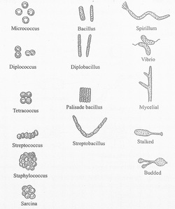
Fig. 2.3 Different forms of bacteria
3 Spirilla (sing., spirillum) : They are cells which are twisted like a screw. They occur as y single cells. E.g., Spirochaete.
4 Vibrio | the cells are curved or comma shaped E.g., Vibrio cholerae.
5 Mycelial or filamentous | They are extensive chains of rod like cells. E.g. Actinomyces
Flagellation in bacteria
Bacterial cells without flagella are called atrichous and those with a flagellum or more flagella art called trichous bacteria.
The trichous forms could be as in figures 2.9.
1 Monotrichous - A single flagellum is produced by the cell.
2 Amphitrichous - A single flagellum at each end of the cell, the cell has two flagella.
3 Cephalotrichous - A cluster of flagella at one end of the cell.
4 Lophotrichous - A group of flagella at two ends of the cell.
5 Peritrichous - Flagella developed all over the cell.
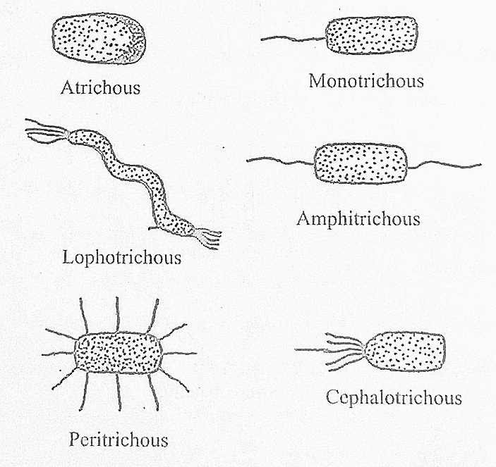
Fig. 2.4 Different kinds of flagellation of bacterial cells
The flagella are made up of a protein called flagellin and they develop from basal granules present in the plasma membrane.
Ultrastructure of bacterial cell
The fine structure seen under high magnification microscopes like electron microscopes is the ultrastructure.
1 Cell wall
The cell wall is chemically made up of peptidoglycans or murein or mucopeptides. It is about 50 to 100 A thick (A = angstroms, 1 A metre). The peptidoglycans are made up of many alternating N-acetylglucosamines and N-acetyl muramic acids which form many chains and these chains are interlocked by tetrapeptides.
In gram positive bacteria the cell wall has upto 90% of peptidoglycans. In gram negative bacteria only 10% of cell wall is peptidoglycan and 90% is the lipopolysaccharide external to cell wall.
2 Slime and capsule
The cell secretes slime made up of polysaccharides, it is a gelatinous sheath around the cell wall. When slime has nitrogenous compounds like amino acids it is called capsule. The slime protects bacteria from dehydration and the capsule protects it from the anti-bodies and phagocytosis by host cells.
3 Plasma membrane
It is present inner to the cell wall. It is a selectively permeable membrane made up of phospholipids and proteins. It has respiratory enzymes and enzymes for DNA replication.
The membranes have the following structures.
I. Mesosomes - These are the invaginations of the membranes. Larger septal mesosomes occur near centre of the ceil and smaller lateral mesosomes away from the centre.
Septal mesosomes help in cell division and lateral mesosomes assist in the reproduction of bacterial DNA or chromosome.
II. Flagella - They develop from basal bodies located in the membrane and are made up of a protein called flagellin.
The cytoplasm is a rich fluid, it is of a viscous consistency and contains carbohydrates, proteins, lipids, enzymes and co-enzymes. There are numerous ribosomes of 50S and 30S subunits. Membrane bound organelles like mitochondria, Golgi complex, lysosomes, etc., are absent. Different types of ribonucleic acid molecules (RNA) are present. There are polyribosomes or polysomes where numerous ribosomal complexes are present attached to messenger RNA (mRNA). From polysomes numerous copies of same protein is made simultaneously and rapidly.
5 Nucleoid or genophore or incipient nucleus
Nucleus is absent. The genetic material DNA is present in a region called genophore. The bacterial chromosome is naked DNA (Deoxyribonucleic acid), since the DNA is not associated with proteins like histones.
6 Plasmids
In addition to the large circular bacterial DNA, there are small sized circular DNA molecules called plasmids in the cytoplasm. The plasmid replicates independently of the main DNA. Plasmids carry many genes for different functions like genes for antibiotic resistance, nif or nitrogen fixation genes and the fertility plasmid (which confers male status to bacterial cells) and such cells have sex pili or conjugation tubes to transfer DNA segments to female cells.
Ultrastructure of bacterial cell
The fine structure seen under high magnification microscopes like electron microscopes is the ultrastructure.
1 Cell wall
The cell wall is chemically made up of peptidoglycans or murein or mucopeptides. It is about 50 to 100 A thick (A = angstroms, 1 A metre). The peptidoglycans are made up of many alternating N-acetylglucosamines and N-acetyl muramic acids which form many chains and these chains are interlocked by tetrapeptides.
In gram positive bacteria the cell wall has upto 90% of peptidoglycans. In gram negative bacteria only 10% of cell wall is peptidoglycan and 90% is the lipopolysaccharide external to cell wall.
2 Slime and capsule
The cell secretes slime made up of polysaccharides, it is a gelatinous sheath around the cell wall. When slime has nitrogenous compounds like amino acids it is called capsule. The slime protects bacteria from dehydration and the capsule protects it from the anti-bodies and phagocytosis by host cells.
3 Plasma membrane
It is present inner to the cell wall. It is a selectively permeable membrane made up of phospholipids and proteins. It has respiratory enzymes and enzymes for DNA replication.
The membranes have the following structures.
I. Mesosomes - These are the invaginations of the membranes. Larger septal mesosomes occur near centre of the ceil and smaller lateral mesosomes away from the centre.
Septal mesosomes help in cell division and lateral mesosomes assist in the reproduction of bacterial DNA or chromosome.
II. Flagella - They develop from basal bodies located in the membrane and are made up of a protein called flagellin.
- Pill - They are also called fimbriae. These hollow structures are made up of pilin protein and are shorter, straighter and thicker than flagella. They help in anchoring the cells in a favourable medium. The sex pili seen only in male cells helps in conjugation process of sexual reproduction.
The cytoplasm is a rich fluid, it is of a viscous consistency and contains carbohydrates, proteins, lipids, enzymes and co-enzymes. There are numerous ribosomes of 50S and 30S subunits. Membrane bound organelles like mitochondria, Golgi complex, lysosomes, etc., are absent. Different types of ribonucleic acid molecules (RNA) are present. There are polyribosomes or polysomes where numerous ribosomal complexes are present attached to messenger RNA (mRNA). From polysomes numerous copies of same protein is made simultaneously and rapidly.
5 Nucleoid or genophore or incipient nucleus
Nucleus is absent. The genetic material DNA is present in a region called genophore. The bacterial chromosome is naked DNA (Deoxyribonucleic acid), since the DNA is not associated with proteins like histones.
6 Plasmids
In addition to the large circular bacterial DNA, there are small sized circular DNA molecules called plasmids in the cytoplasm. The plasmid replicates independently of the main DNA. Plasmids carry many genes for different functions like genes for antibiotic resistance, nif or nitrogen fixation genes and the fertility plasmid (which confers male status to bacterial cells) and such cells have sex pili or conjugation tubes to transfer DNA segments to female cells.
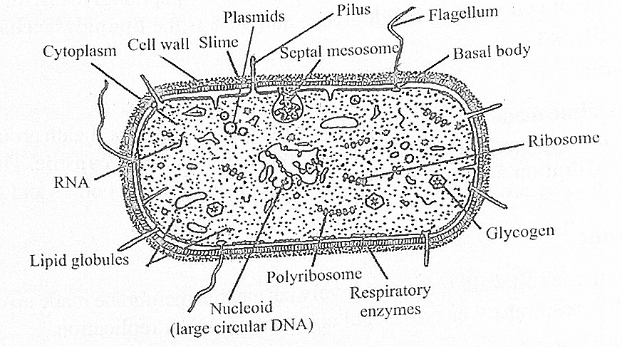
Fig. 2.10 Ultrastructure of a bacterial cell
Nutrition in bacteria
Bacteria show autotrophic and heterotrophic modes of nutrition.
Autotrophic bacteria : They produce organic food or nutrients from simple inorganic substances. They are of two types namely
1. Photosynthetic and
2. Chemosynthetic.
Heterotrophic nutrition
The bacteria get readymade organic food from different sources. Based upon the nature of the source, heterotrophs could be saprophytes, parasities or symbionts.
1 Saprophytes : These bacteria are also called decomposers, transformers, detrivores (dirt eaters) or osmotrophs.They obtain organic food by decomposing dead bodies and excreta of animals and dead plant parts like leaves. These bacterial cells produce enzymes that come out of the cell (extra cellulai enzymes) and breakdown complex molecules into simple ones which enter the cell. From these simplemolecules the cell can synthesise the required complex molecules. Some of the extracellular enzymes are cellulases, proteases and lipases which breakdown celluloses, proteins and lipids respectively into j simple molecules.
2 Parasites : Parasitic bacteria attack living plants and animals and obtain organic nutrients from them. These bacteria cause diseases and such parasites are called pathogens (disease causers). As an example we have Salmonella typhimurium\ this bacteria lives in our intestines and gets organic food and the bacteria secretes toxins for typhoid fever in humans.
3 Symbionts : As symbiotic bacteria like Rhizobium spp and Bacillus spp cause nodule formation in legume plant roots. These bacteria generate useful nitrate compounds for the legume plants and the legume plant is a source of organic food like sugars for the bacteria. This mutually beneficial relationship j between two types of organisms is called symbiosis.
Respiration in bacteria
Bacterial respiration is of two types namely aerobic or oxygen requiring and anaerobic or not needing oxygen. Respiration is an energy creating process where organic food is broken down and the energy released is used to make high energy biomolecules like Adenosine tri phosphate (ATP) and these high energy biomolecules are used to synthesise various biomolecules required by the cell.
1 Bacteria with aerobic process are called aerobes and those with anaerobic process are called anaerobes. Aerobes and anaerobes could be obligate or facultative.
2 Obligate aerobes - They compulsorily require oxygen to survive, e.g., Bacillus subtilis.
3 Obligate anaerobes - They grow in places where oxygen is absent like deep sewers, e.g., Clostridium botulinum.
4 Facultative aerobes - Most of the photosynthetic bacteria are able to live (or respire) with or without oxygen.
5 Facultative anaerobes - They show aerobic process but if there is temporary absence of oxygen they take up the anaerobic process.
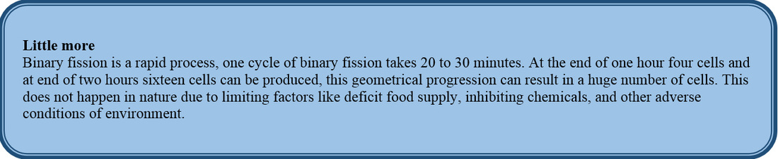
Reproduction in bacteria
Bacteria reproduce by asexual and sexual (parasexual) processes; asexual reproduction occurs by binary fission and endospore formation.
Binary fission: It is a simple type of cell division wherein the cellular contents are divided into two parts after the replication or reproduction of main circular chromosomal DNA. The lateral mesosomes assist in DNA reproduction, mesosomes play a role in positioning of the two double stranded DNA to opposite zones of the cell. The mesosomes also play a role in distribution or sequestration of cellular components to two parts of the cell (the future daughter cells). A constriction appears at the centre of the cell, the septal mesosomes are involved, the constriction deepens and grows centripetally from margin to centre and finally two cells are produced.
Bacteria show autotrophic and heterotrophic modes of nutrition.
Autotrophic bacteria : They produce organic food or nutrients from simple inorganic substances. They are of two types namely
1. Photosynthetic and
2. Chemosynthetic.
Heterotrophic nutrition
The bacteria get readymade organic food from different sources. Based upon the nature of the source, heterotrophs could be saprophytes, parasities or symbionts.
1 Saprophytes : These bacteria are also called decomposers, transformers, detrivores (dirt eaters) or osmotrophs.They obtain organic food by decomposing dead bodies and excreta of animals and dead plant parts like leaves. These bacterial cells produce enzymes that come out of the cell (extra cellulai enzymes) and breakdown complex molecules into simple ones which enter the cell. From these simplemolecules the cell can synthesise the required complex molecules. Some of the extracellular enzymes are cellulases, proteases and lipases which breakdown celluloses, proteins and lipids respectively into j simple molecules.
2 Parasites : Parasitic bacteria attack living plants and animals and obtain organic nutrients from them. These bacteria cause diseases and such parasites are called pathogens (disease causers). As an example we have Salmonella typhimurium\ this bacteria lives in our intestines and gets organic food and the bacteria secretes toxins for typhoid fever in humans.
3 Symbionts : As symbiotic bacteria like Rhizobium spp and Bacillus spp cause nodule formation in legume plant roots. These bacteria generate useful nitrate compounds for the legume plants and the legume plant is a source of organic food like sugars for the bacteria. This mutually beneficial relationship j between two types of organisms is called symbiosis.
Respiration in bacteria
Bacterial respiration is of two types namely aerobic or oxygen requiring and anaerobic or not needing oxygen. Respiration is an energy creating process where organic food is broken down and the energy released is used to make high energy biomolecules like Adenosine tri phosphate (ATP) and these high energy biomolecules are used to synthesise various biomolecules required by the cell.
1 Bacteria with aerobic process are called aerobes and those with anaerobic process are called anaerobes. Aerobes and anaerobes could be obligate or facultative.
2 Obligate aerobes - They compulsorily require oxygen to survive, e.g., Bacillus subtilis.
3 Obligate anaerobes - They grow in places where oxygen is absent like deep sewers, e.g., Clostridium botulinum.
4 Facultative aerobes - Most of the photosynthetic bacteria are able to live (or respire) with or without oxygen.
5 Facultative anaerobes - They show aerobic process but if there is temporary absence of oxygen they take up the anaerobic process.
Reproduction in bacteria
Bacteria reproduce by asexual and sexual (parasexual) processes; asexual reproduction occurs by binary fission and endospore formation.
Binary fission: It is a simple type of cell division wherein the cellular contents are divided into two parts after the replication or reproduction of main circular chromosomal DNA. The lateral mesosomes assist in DNA reproduction, mesosomes play a role in positioning of the two double stranded DNA to opposite zones of the cell. The mesosomes also play a role in distribution or sequestration of cellular components to two parts of the cell (the future daughter cells). A constriction appears at the centre of the cell, the septal mesosomes are involved, the constriction deepens and grows centripetally from margin to centre and finally two cells are produced.
Endospore formation
The process of endospore formation is called sporulation. These spores can resist unfavourable environmental conditions like dehydration, nutrient absence and high temperatures.
Endospores are perennatory structures which help in survival. Cells that cannot form endospores could die during unfavourable conditions.
Endospores are commonly produced by bacteria belonging to the genera Clostridium and Bacillus. One cell forms only one endospore, and with the return of favourable environmental conditions the endospore generates a cell that can reproduce by usual methods. Endospores also help in dispersal of bacterial cell. Endospores remain viable for many years.
The endospore has many wall layers like the outermost exosporium and it is followed by spore coat of specific proteins, the spore coat encloses the cortex which has the peptidoglycan structure with tetrapeptide cross linkages. The endospore wall has heat resistant chemicals like sialic acid and dipicolinic acid.
Sexual reproduction (parasexual) in bacteria
Sexual reproduction takes place by a parasexual process. In bacteria, one of the aims of sexual process is to achieve genetic recombination and bacteria achieve this by three methods namely transformation, transduction and conjugation.
Transformation: It is process where segments of DNA (genes) are transferred from one bacterial cells (very often dead cells) to another living cell via the liquid medium. The process was discovered by Griffith in 1928. In his experiments he used two strains of pneumonia causing bacteria Streptococcus pneumoniae. There are two strains of this bacteria namely virulent strains with a capsule that can cause disease and harmless avirulent strain without capsule (Formation of capsule is a genetic character requiring the necessary genes or DNA). In one of his experiments Griffith mixed avirulent living strain and heat killed virulent strain and when the mixture was injected into mice the mice died. Griffith concluded that a transforming principle entered the living noncapsulated avirulent cells and made them virulent (they made a capsule). The transforming principle was later on identified as DNA (the genetic material) and this acquiring a new character (capsule formation) is an example of genetic recombination.
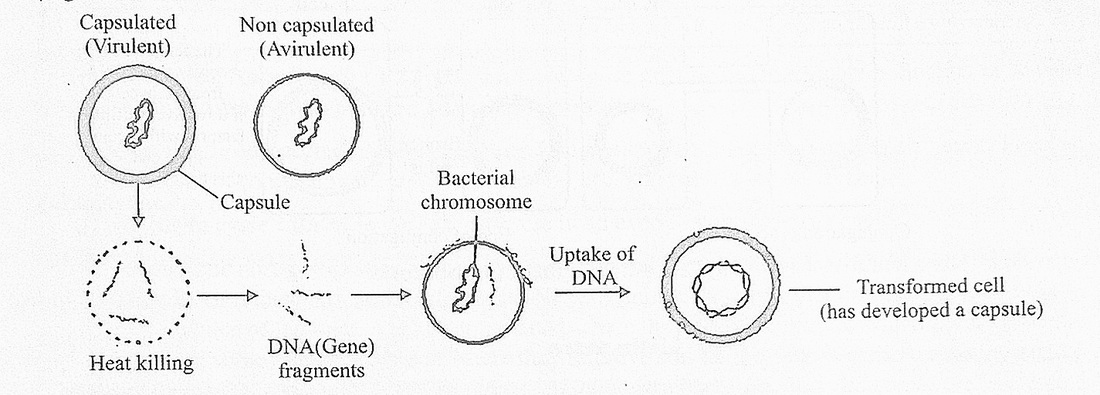
Fig.-2.11 Transformation in bacteria
Transduction: In this process segment of DNA (genes) are transferred from one bacterium to another by the agency of viruses (bacteriophages) and the bacteria that received the gene acquires a new character resulting in genetic recombination. The process of transduction was discovered in the bacterium Salmonella typhimurium by Zinder and Lederberg in 1952.
Conjugation: The process was discovered by Lederberg and Tatum in 1946 in Escherichia coli strain K12. The male cell (donor cells) has fertility plasmid or F -factor and male cell produces 2 to 5 sex pili per cell which function as conjugation tubes, which connect itself to cell wall of female cells (recipient cells) which to not have fertility factors. Through the conjugation tube chromosomal DNA is transferred from male to female cells, only a small part of DNA can be transferred because conjugation occurs for a short time period. The fertility plasmid often integrates with the main bacterial chromosome and then it is called an episome and such cells are called High frequency recombinants (Hfr) cells, since these cells can transfer genes at a very high frequency or rate.
During conjugation if the F+ plasmid DNA enters female cell the female cell becomes male and can function as a donor in future conjugation process.
Economic importance of bacteria
Bacteria have numerous beneficial uses economically and also they cause a lot of damage in the form of food spoilage and causing diseases in plants, animals and humans. They are friends and foes of human beings.
Beneficial activities
a) Natural scavengers: Bacteria show saprophytic nutrition during which they decompose deal bodies of plants and animals and animal excreta. During this process the bacterial cells secrete extra cellular enzymes that come out of the cell and breakdown complex molecules into simple ones which are absorbed as food or nutrient by the cell. The bacterium Cytophaga can decompose polysaccharides like cellulose into monosaccharides which are absorbed by the cell. Since bacteria clean up the environment by decomposing activities they are called natural scavengers.
b) Fermentation: This is a type of anaerobic respiration performed by bacteria. Clostridium spec ferments sugars into butyric acid, Acetobacter aceti is used in vinegar manufacture, bade fermentation is used in cheese and yogurt making.
c) Retting : The fibrous tissues of plants like coconut and jute are immersed in water ar bacteria in water hydrolyses the middle lamella of pectic substances joining the individual The fibers get separated and then are used in making ropes or gunny bags.
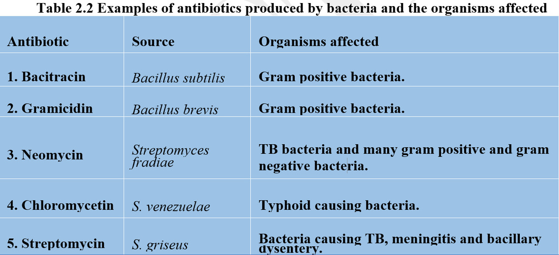
d) Antibiotic production: Antibiotics are secondary metabolites produced by bacterial cel1 can destroy other microbes. The genus Streptomyces has many species that produce antibiotics.
a) Natural scavengers: Bacteria show saprophytic nutrition during which they decompose deal bodies of plants and animals and animal excreta. During this process the bacterial cells secrete extra cellular enzymes that come out of the cell and breakdown complex molecules into simple ones which are absorbed as food or nutrient by the cell. The bacterium Cytophaga can decompose polysaccharides like cellulose into monosaccharides which are absorbed by the cell. Since bacteria clean up the environment by decomposing activities they are called natural scavengers.
b) Fermentation: This is a type of anaerobic respiration performed by bacteria. Clostridium spec ferments sugars into butyric acid, Acetobacter aceti is used in vinegar manufacture, bade fermentation is used in cheese and yogurt making.
c) Retting : The fibrous tissues of plants like coconut and jute are immersed in water ar bacteria in water hydrolyses the middle lamella of pectic substances joining the individual The fibers get separated and then are used in making ropes or gunny bags.
d) Antibiotic production: Antibiotics are secondary metabolites produced by bacterial cel1 can destroy other microbes. The genus Streptomyces has many species that produce antibiotics.
e) Ecological importance: Bacteria can decompose organic biodegradable pollutants in sewage water and are used in purification of sewage water.Genetically engineered bacteria like Psuedomonas spp can decompose crude petroleum. This can be useful in controlling oil spills in oceans by accidents involving oil tankers.
f) Nitrogen cycle: Bacteria play a major role in all events of the nitrogen cycle. Asymbiotic nitrogen fixation is done by free living soil bacteria like Clostridium and Azotobacter. They fix or reduce nitrogen to ammonia (NH3). Symbiotic bacteria that fix nitrogen are found in root nodules of legumes (angiosperm plants) where they generate nitrates which are used by the legumes; some nitrates enter soil where they improve soil fertility, the bacteria benefit by getting organic food from the plants, Rhizobium leguminosarum and Bacillus radicicola are associated in nodules.
Bacteria like Nitrosomonas oxidise ammonium (NH4+) compounds into nitrites (N02_) and bacteria like Nitrosobacter convert nitrites into nitrates (N03~)
Another important event of nitrogen cycle is the process of denitrification where free nitrogen is released from N03 and NH3. Denitrification is brought about by bacteria like Pseudomonas, Thiobacillus denitrificans and Bacillus subtilis.
g) Importance in genetic engineering: Bacterial cells of Escherichia coli have been made to produce pure grade human insulin (humulin) by incorporating insulin gene into plasmids and then introducing the recombinant plasmids (plasmids with foreign genes like insulin gene and human growth hormone gene) into the bacterial cells and these bacterial cells in the culture medium produce the gene products which are used by human beings. Bacteria like Agrobacterium tumefaciens have Ti or tumor inducing plasmids. These plasmids are extensively used in genetic engineering or biotechnology.
h) Extraction of minerals from ores: Species of bacteria.belonging to genera like Sulfobolus and Thiobacillus are used is microbial leaching or extraction of useful elements like copper from low grade copper ores.
Harmful bacteria
Bacteria are responsible for many harmful activities like food spoilage, food poisoning and as pathogens of numerous plant, animal and human diseases.
Food spoilage: Some saprophytic bacteria spoil food by changing the appearance of food and causing unpleasant aroma. Species of bacteria belonging to genera like Lactobacillus and Streptobacillus spoil milk and milk products like butter and cheese.
Food poisoning: Clostridium botulinum bacteria commonly produces toxins in improperly canned dnned food, they generate toxins that cause fatal problem of botulism which can kill humans by causing respiratory paralysis. Symptoms of botulism are swelling of the tongue and double vision.
Salmonellosis is an infection of eggs and poultry by Salmonella bacteria that can cause fever and typhoid infection.
Plant diseases caused by bacteria
Bacteria cause numerous plant diseases. The most common is citrus canker caused mainly in lemon leaves and fruits. It is caused by the bacterium Xanthomonas citri; necrotic (tissue damaging) lesions develop round green raised spots. Later the spots become brownish / grey, rupture and develop a hard corky tissue which makes lemon fruits unattractive.
High moisture content and wind plays a role in spread of canker disease.
Isolation and burning down of infected plants or spray of copper compounds can control the disease.
Erwinia carotovora causes soft rot is carrot plants where the edible roots are damaged.
Xanthomonas oryzae causes blight disease in rice plants.
Agrobacterium tumefaciens causes crown gall disease in apple trees and rose plants.
Human and animal diseases caused by bacteria
Anthrax: In addition to humans, anthrax is a disease of horses, cattle and sheep caused by gram positive bacillus Anthracis. They enter the host body by means of spores. Protection is by vaccination of live avirulent bacilli. Penicillin given in large doses at early stage can cure humans of anthrax.
Cholera: The gram negative Vibrio cholerae infects the gastrointestinal tract. It causes severe diarrhoea in humans.
Consumption of sewage contaminated water or food is the cause.
The patient is dehydrated, therefore the patient should have oral rehydration therapy (ORT) where they drink lots of salt and sugar containing water. The antibiotic tetracycline can control the bacteria.
Gastric ulcer: This problem and gastritis are caused by a gram negative spiral, Jbacteria Helicobacter pylori. The bacteria produce the urease enzyme which enables the bacteria to survive in the acidic environment of gastric mucosa.
Tuberculosis: The bacterium Mycobacterium tuberculosis attacks lungs and causes necrotic lesions where lung tissues are destroyed. Immunisation is by vaccination with a vaccine containing live attenuated cells of bacteria. The vaccine is called BCG (Bacille Calmette Guerin)
Sexually transmitted diseases (STD): Gonorrhea caused by Neisseria gonorrhoeae and syphilis caused by Treponema pallidum are the two common STDs, also called veneral disease. They are caused by sexual intercourse with infected persons.
Syphilis has a primary stage that expresses in the patient 10- 90 days after infection during which sores appear in external genitalia of men and women and swelling of local lymph nodes, if not treated with antibiotics the disease enters secondary and tertiary stages where even the nervous system is affected.
Penicillin and other antibiotics are used in treatment of STDs.
2.3.2 Cyanobacteria
These monerans (belong to Division Cyanophyta) were earlier called blue green algae. They have many characters similar to bacteria, hence the term cyanobacteria (They also differ from bacteria in some characters).
Some common examples of cyanobacteria are Nostoc, Spriulina, Anabaena and Rivularia. They have adapted themselves to grow in a wide variety of habitats. They occur in hot springs where temperature as high as 80°c+ is common, they are present in fresh and sea water. In polluted waters they grow to a huge population causing water blooms; they also grow on moist soils.
Cyanobacteria occur as
I. Single cells as in Chroococcus.
II. Colonies as in Microcystis and Gloeocapsa.
III. Filaments as in Nostoc, Oscillatoria and Anabaena.
There are about 150 genera and 1500 species of blue green algae.
All of them are photosynthetic with different types of light capturing pigments. The green pigments are chlorophyll a, blue pigment is c-phycocyanin, red pigment is c-phycoerythrin yellow pigments are xanthophylls. Cell structure is typically prokaryotic. Flagella and sexual reproduction are absent.
Vegetative reproduction is by fragmentation of thallus (simple plant body without tissues) or by hormogonia formation.
Special cells called akinetes and heterocysts also help asexual reproduction.
Nostoc is a commonly found blue green alga and its systematic position is
Kingdom: Monera
Division : Cyanophyta (Cyanobacteria)
Class : Cyanophyceae
Order : Nostocales (Hormogoniales)
Family : Nostocaceae
Genes : Nostoc
The genus has about 30 species and is very common is paddy fields, some are phytoplankton, some species grow as symbionts in gametophytes of bryophytes like Anthoceros, some live in coralloid roots of the gymnosperm, Cycas Nostoc occur in nature as mucilaginous spheres and each sphere has numerous filaments of Nostoc, the hydrophilic chemical mucilage is secreted by the cells. The mucilage retains water and protects the thallus from getting dehydrated when the environment is almost dry. The mucilaginous masses range in size from 20 to 60 mm.
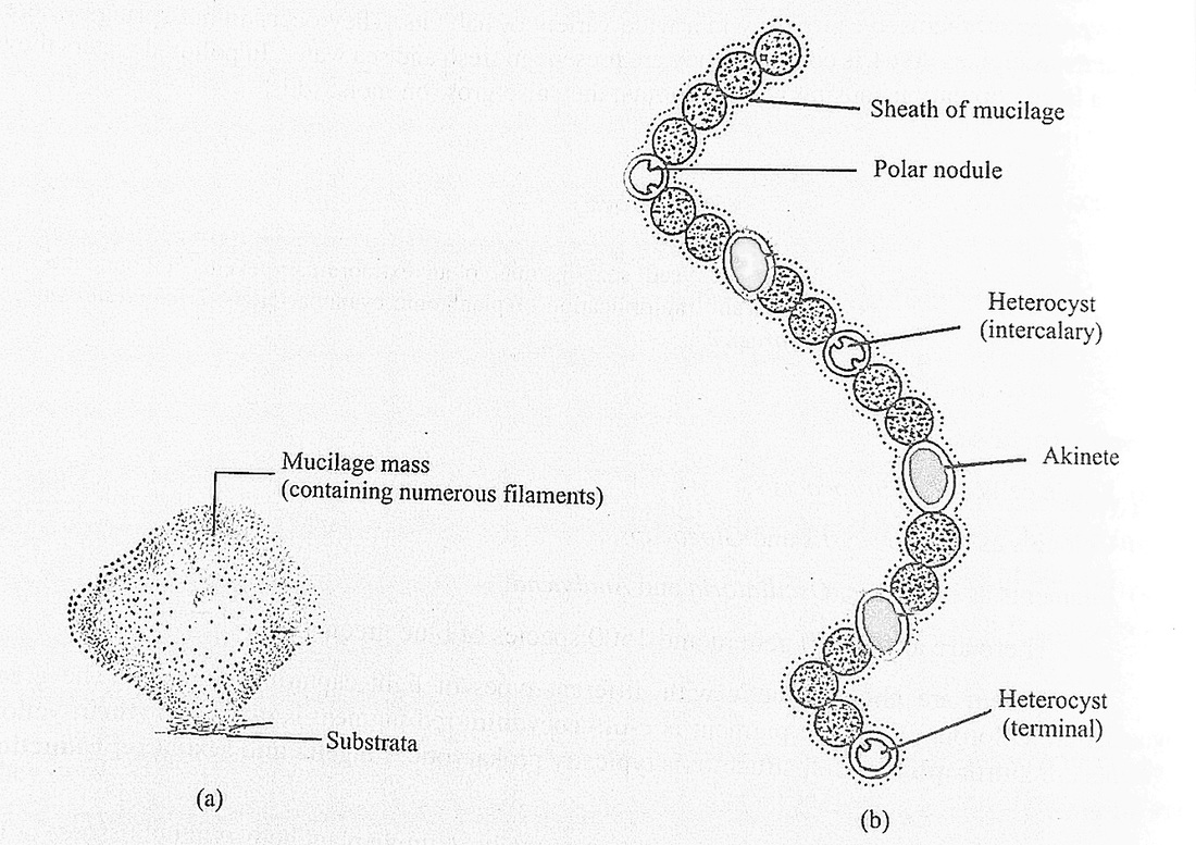
Fig.-12 (a)Nostoc colony (b) Filamentuos structure
Structure of trichome
The trichome is unbranched and is a chain of oval or spherical cells. The cells secrete the mucilage sheath and the trichome with the sheath is a filament (In Nostoc the filament has one trichome so it is called a monotrichomic filament). In a trichome most of the cells are vegetative and have similar structure.
Some cells are modified as heterocysts, they are hyaline, may be polar or intercalary, polar heterocyst have one polar pore when young, in old heterocyst the pore is blocked by a polar nodule. The intercalary heterocyst has two polar pores and two polar nodules. They help in reproduction by hormogonia formation and they have nitrogenase enzyme which is responsible for nitrogen fixation, where molecular nitrogen is oxidised to nitrates which increases soil fertility.
Some of the vegetative cells also enlarge slightly (by storing more chemical products) and develop a thick wall, these cells are called akinetes. They help in perennation (survival) during unfavourable conditions and they are resting cells. They also help in reproduction when environmental conditions become favourable.
Ultrastructure of the cell
The spherical or oval cells are bounded by a peptidoglycan cell wall, surrounded by a mucilage sheath. Within the cell wall is a plasma membrane. The peripheral part of the cell is the coloured chromoplasm and the central colourless centroplasm where the genetic material DNA is present.
In the chromoplasm there are numerous membrane systems called lamellae or thylakoids, the photosynthetic pigments (chlorophyll a, xanthophylls like the unique myxoxanthin) are associated with these membranes. The chromoplasm has reserve food materials like cyanophycean starch, oil drops. The ribosomes are of 50S and 30S types which associate to form 70S complexes during protein synthesis. All membrane bound organelles like true vacuoles, mitochondria, plastids, endoplasmic reticulum and Golgi bodies are absent.
The chromoplasm encloses the central colourless centroplasm where the naked DNA is present. From the DNA different types of RNA molecules are produced like messenger RNA, transfer RNA and ribosomal RNA which are necessary for protein synthesis. The centroplasm represents a primitive nuclear zone (nuclear membrane and nucleoli are absent) and is also called nucleoid, genophore or incipient nucleus.
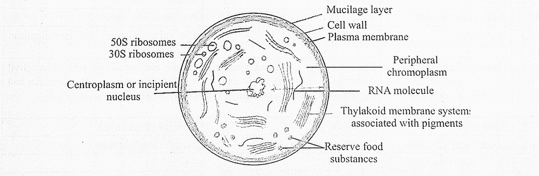
Reproduction
In Nostoc sexual reproduction is absent the asexual methods of reproduction are as follows:
a) Fragmentation: When the mucilaginous thallus grows to a large size it breaks apart (by natural disturbances like water currents, biting of fishes) into numerous thalli which grow to bigger size.
b) Hormogonia or Hormogone formation : Hormogonia are small segments of a filament, the filament often break at the point of heterocysts which probably are the weak links of the filament. The hormogonia come out and grow into larger filaments and develop new thalli which get surrounded by mucilaginous mass secreted by the cells.
c) Akinetes : These cells are produced when water becomes scarce, some cells at random store food and develop thick wall to form structures for survival (perennation) called akinetes. The cells that cannot form akinetes could die. When conditions become favourable the contents of the akinete develop into Nostoc filaments which come out and grow.
d) Heterocysts : In Nostoc commune the contents of the heterocyst divide to form a number of spores, which by cell division form new trichomes
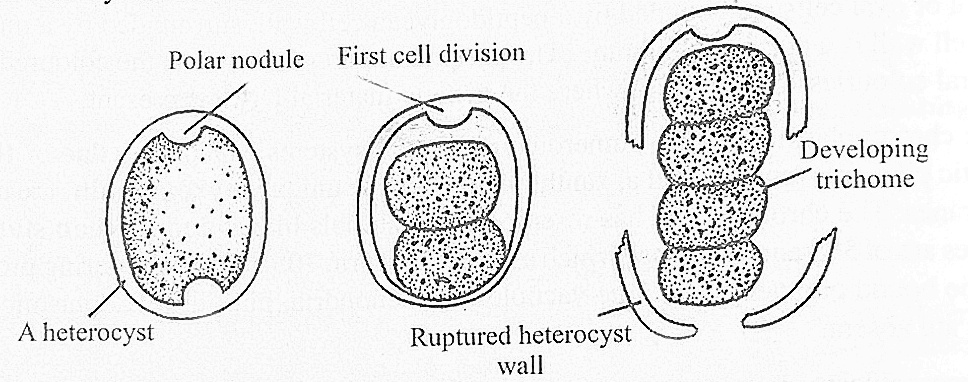
Fig.-2.13 Reproduction in heterocysts
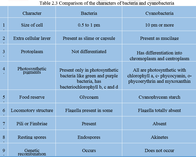
Economic importance of cyanobacteria Useful cyanobacteria
1) The aquatic forms are the food producers and they form the base of the food pyramid. Primary consumers directly depend on producers for food (energy) source.
2 Some cyanobacteria have heterocysts and all such blue green algae function as biofertilisers because these soil forms fix nitrogen to useful forms like nitrates, thereby improving soil fertility. Some of the nitrogen fixers are Nostoc, Tolypothrix and Anabaena.
3 The mucilage found is soil cyanobacteria keeps the soil particles together, thereby preventing soil erosion. Anabaena is an effective soil binder.
4 Some like Spirulina are being cultivated for their numerous uses.
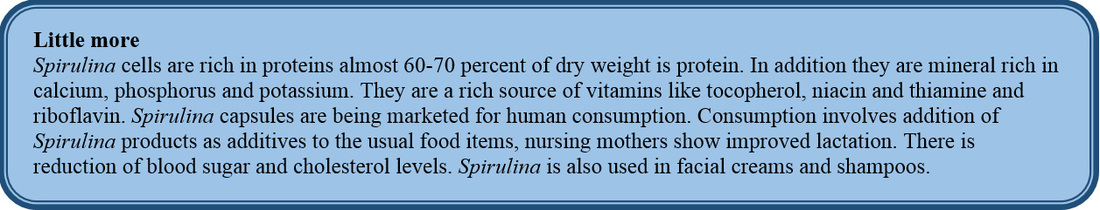
5 Extracts of Anabaena is used in biological control (the process where an unwanted organism is removed by using another organism or its chemical product) of mosquito larvae, the larvae are killed by the extracts. Biocontrol protects the environment from pollution by chemical insecticides.
Harmful cyanobacteria
1. Some cyanobacteria produce toxins, for example Anabaena lemmermani produces toxins that can kill fishes and even cattle.
2. In fresh water ponds and lakes which are polluted by nitrogenous compounds like fertilisers (washed into the lake from surrounding agricultural fields) the blue green algae like Microcystis grow in large numbers, they create water blooms, such water emits unpleasant odour and is unfit for human use.
3. The water blooms use up the oxygen in water at night thereby killing the fishes and other aerobic aquatic organisms. The dead bodies of these organisms decompose to pollute the water
2.3.3 Rickettsias
H.T. Ricketts discovered these monerans in 1909 while investigating Rocky Mountain spotted fever. Rickettsias are smaller than bacteria with a smaller number of genes. They are rod or sphere I shaped unicells, reproducing only by binary fission, they are pathogens of diseases like scrub fever and I typhus fever.
2.3.4 Actinomycetes
These monerans are bacteria like cells forming radiating filaments of cells. They have a combination \ of bacterial and true fungal characters. They are gram +ve and grow as radiating colonies in the culture 1 media hence the name actinomycetes (actino= star like; mycetes =fungi) (ray fungi) They grow as 1 saprophytes on substrata rich in dead organic matter. They resemble bacteria in their physiology and are referred to as evolved bacteria.
Actinomycetes belonging to genus Streptomyces produce many antibiotics which are used to 1 control many bacteria caused diseases in humans.
S. griseus is the source of antibiotic streptomycin.
S. venezuelae is the source of antibiotic Chloromycetin.
S. rimosus is the source of antibotic terramycin.
S. fradiae is the source of antibiotic neomycin.
2.3.5 Mycoplasma (pleuropneumonia like organisms or PPLO)
These monerans belong to the Class Mollicutes. They were discovered by Nocard and Roux in 1898 from lung fluids of cattle affected by bovine pleuropneumonia. The cells do not have cell wall and are 0.1 to 0.15 pm in size. The plasma membrane is the outermost cellular boundary, the cell changes shape very often. Like all prokaryotes, they have no nucleus and membrane bound organelles like j mitochondria, plastids, true vacuoles, Golgi bodies, lysosomes, cytoskeletal structures are also absent Ribosomes are of 70S types. Cytoplasm has many granules. A double stranded circular DNA is found in the cytoplasm. Cells do not have flagella. Enzymes are bound to plasma membrane and some enzymes occur in cytosol (cytoplasm)
Mycoplasmas generally lead a parasitic existence in animals and plants.
Some of the plant diseases caused by mycoplasma are potato purple dwarf, dwarf disease in mulberry plants, witches broom in legume plants and papaya plant bunchy top disease.
They cause pleuropneumonia in humans and cattle.
2.3.6 Archaebacterla
The members of this domain are called Archaea (Gr., archaio = ancient).
These organisms have the following characters
1) They are the most ancient or most primitive prokaryotes.
2) They are found in high stress environment of high salt content (salt basins), high temperature zones in hot springs and oceanic thermal vents.
3) The cellular plasma membrane has special lipids to withstand the stresses of high temperature.The lipids of archea have glycerol linked to branched chain hydrocarbons, whereas in bacteria glycerol is linked to fatty acids.
4) The cell walls do not have peptidoglycan and cellulose. Cell wall is made up of proteins and non- cellulosic polysaccharides.
5) Most of them are chemoautotrophs. There are no parasites (since the environment does not have hosts)
6) Archaea share numerous features with eukaryotes in having similar types of ribosomal proteins, simliar types of transcription process (where DNA produces different types of RNA). They also have similar types of transfer RNA molecules like the eukaryotes.
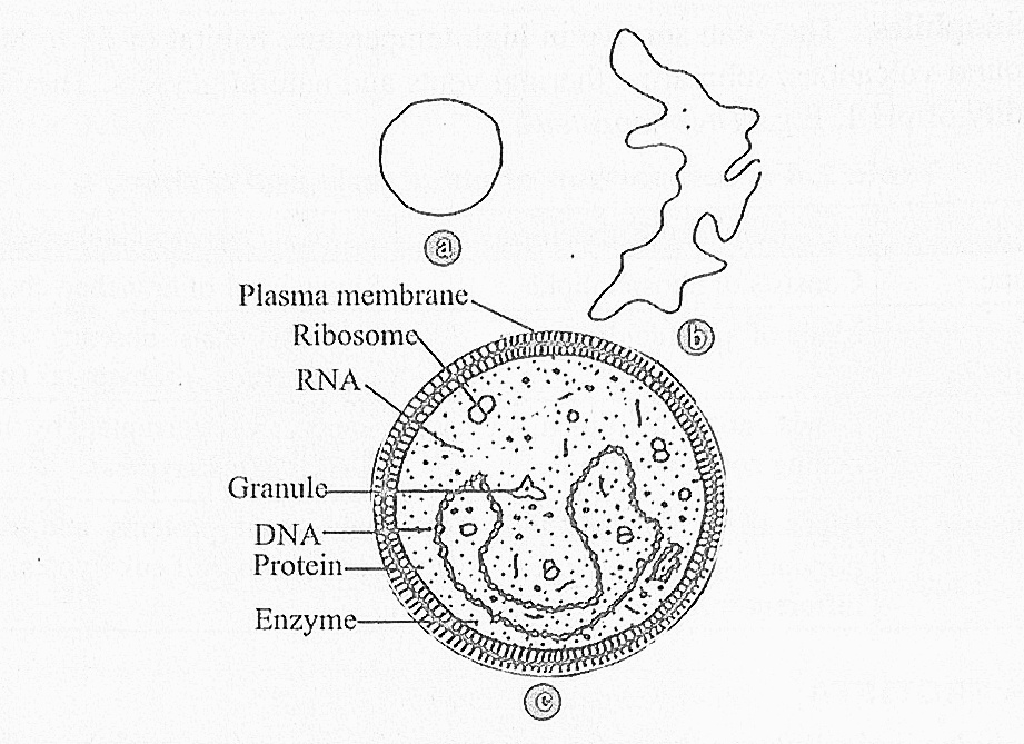
Fig. 2.16 Structure of mycoplasma: (a) spherical shape
(b) branched filamentous shape (c) details of cell structure
(b) branched filamentous shape (c) details of cell structure
Archaebacteria are of the following three types
1) Methanogens: They produce almost 65% of atmospheric methane. They live in anaerobic environment like swamps, deep sewage, human colon, rumen of cattle. These forms of archaea are used in biogas generators. E.g., Methanococcus.
2) Halophiles: They are the archaea of salty habitats. They are mostly anaerobes. They have a chemical - halorhodopsin to pump in chlorides into the cell to prevent cellular dehydration. They also have chemicals to synthesise ATP in presence of light. E.g., Halobacterium.
3) Thermoacidophiles: They can survive in high temperature habitat of 65 to 80°C like sulphur springs, around volcanoes, submarine thermal vents and natural geysers. They can also survive at high acidity of pH 1. E.g., Thennoproteus.
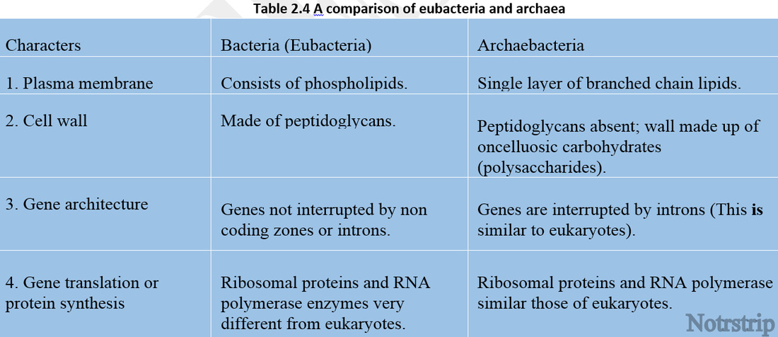
2.4 KINGDOM PROTISTA
The kingdom includes unicellular eukaryotic, microscopic organisms which mostly are aquatic. It includes organisms called protists. The protista is a phylogenetic or evolutionary link between prokaryotic monera and multicellular kingdoms like Plantae, Mycota and Animalia.
Some protists like Euglena are photosynthetic like plants, some protists like slime moulds are saprophytic like mycotans, some like Didynia are predators like some animalia members^, Many photosynthetic protists like diatoms are free living fresh or sea water microscopic organisms called phytoplankton. They have chlorophyll and are capable of photosynthesis. The different types of phytoplankton are the major food producers of the oceans and they support different types of consumers directly and indirectly as a food or energy source.
Structure of a typical protist
All protists have a typical eukaryotic cell structure with the presence of a true nucleus with nuclear membrane and nucleoli. Membrane bound organelles like true vacuoles, mitochondrial Bplastids. lysosomes, endoplasmic reticulum are present. Photosynthetic forms have chloroplasts but no cell wall as in Euglena. In diatoms, photosynthetic organelle is the golden brown coloured chrommophores and diatoms have cell wall.
Locomotion in protists
Locomotion of cells is by flagella, cilia, psuedopodia, wriggling of cells and by mucilage secretion.
Flagella: They are elongated thread like aids for movement in water. Whip like motion of flagella moves the cells at the rate of about 250 pm per second. Flagellate motion is seen in Euglenoids. Dinofiagellates and Zooflagellates. A ceil produces one or few flagella.
Cilia: They are produced in thousands per cell and are much shorter than flagella. They move in a coordinated way to propel the cell 400 to 2000 pm (or 2 mm) per second as in Paramecium.
Psuedopodia: They are protoplasmic extensions of the cell like in Amoeba and slime moulds. Rate of movement is 3 to 4 pm per second. Psuedopodia means false feet, there are different types of pseudopodia.
Lobopodia are broad and blunt.
Filopodia are slender.
Axopodia are slender with an axial filament.
Reticulopodia are slender and branched.
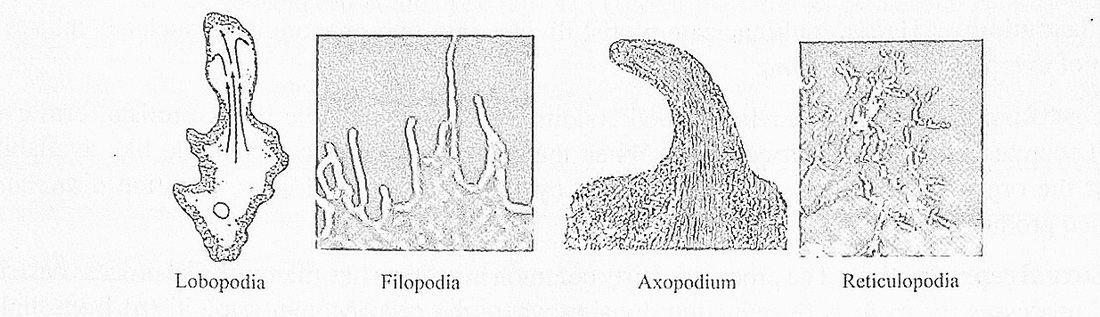
Wriggling: It is a slow wriggling movement caused by expansion and contraction of cell, commonly seen in non-flagellate euglenoids and sporozoans.
Mucilage propulsion: Some protists like diatoms secrete mucilage in one direction and move in opposite direction.
Nutrition in protists
Protista have evolved many types of nutrition (methods to obtain organic food or nutrients is nutrition).
1) Photosynthetic (holophytic): These autotrophic protists can perform photosynthesis since they have chlorophyll, examples are diatoms, euglenoids and dinoflagellates.
2) Holozoic : The organism takes in solid food and digests them in a food vacuole as in Amoeba or food particles are swept into a cellular digestive structure in unicellular protist like Paramecium. This is also called phagotrophic or zootrophic nutrition.
3) Saprophytic : This type of nutrition is shown by decomposers or saprophytes like slime moulds. They live on dead leaves or excreta and secrete enzymes which come out of the cell and digest complex molecules into simple ones which are absorbed by the surface of the body.
4) Parasitic : Parasitic protists like Entamoeba, Trypanosoma, Plasmodium and Girardia get organic nutrients from the body of the host.
5) Myxotrophic : It is a process where an organism can have 2 or more types of nutrition. For example Euglena is holophytic (photosynthetic), saprophytic and holozoic.
Reproduction in protists
They reproduce by asexual and sexual methods.
Asexual reproduction: It is a common type of reproduction in Protista. New individuals produced have the same genetic characters of the parent and are called clones.
Asexual reproduction occurs by fission, plasmotomy and cyst formation.
Fission: Here a cell splits into two daughter cells and each cell gets a nucleus (in the parent cell nucleus divides by mitosis). When a parent cell forms only two cells it is called binary fission Sometimes a parent cell may split into numerous daughter cells and this process is called multiply fission. Euglena divides by binary fission.
Plasmotomy: Here a multinucleate protist divides into two or more multinucleate entities by division of cytoplasm as in Opalina.
Cyst formation: When environmental conditions are unfavourable many protists form cysts. The cell secretes a thick resistant covering. When the conditions become favourable like availability of water the organism comes out of the cyst. The cyst helps is survival or perennation. Entamoeba histolytica produces cysts.
Sexual reproduction: The process is fairly common in protists like diatoms. The process basically I has two processes (a) meiosis or reduction division where the cell becomes haploid (n) from diploid (2n) state. In haploid state the nucleus has one set of chromosomes and in diploid state the nucleus has two sets of chromosomes, During meiosis two events occur; one is reduction of chromosome number by half (50%) and genetic recombination which produces new genetic types of gametes which creates j variations among members of a species, which is essential for organic evolution.
(b) The haploid gametes fuse to form a diploid cell called zygote by the process of syngamyor fertilisation.
Sexual reproduction with meiosis, developed by Protista is an important event in evolutional) biology.
Life cycle of protists
Protists have either haploid or diploid nucleus and meiosis occurs once in their life cycle.
There are two types of life cycles based upon the timing of meiosis.
Life cycle, type I
The protists have haploid nucleus, they reproduce asexually by mitosis or equational division (during I mitosis there no difference in chromosome number of parent and daughter cells).
During sexual reproduce haploid cells fuse (fertilisation) to form diploid zygote. The zygote I undergoes meiosis, to form haploid cells. This process where meiosis occurs in zygote is called zygotic meiosis.
Life cycle, type II
The protists are unicellular with a diploid nucleus. They reproduce asexually by cell division when the 2n nucleus divides mitotically to form two 2n nuclei each of which enters daughter cells.
During sexual reproduction the diploid nucleus undergoes reduction division or meiosis to form haploid nuclei which develop or produce haploid gametes (this is called gametic meiosis since meiosis 1 occurs during gamete formation). The diploid zygote produced after fertilization develops into the 1 diploid unicellular organism, this is seen in Protista like diatoms.
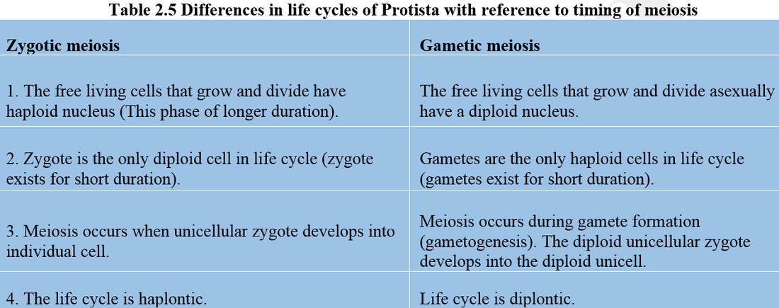
Classification of protista
The protists can be grouped into three major groups.
I. Photosynthetic protists: (organic food producers) they are the protistan algae like Dinoflagellates and Chrysophytes, Euglenoids or Euglenids.
II. Slime moulds: They include saprophytes or decomposers which are food consumers.
III. Protozoan protists: The group includes the protozoans which have different modes of nutrition like holozoic, saprophytic and parasitic.
The group algae have tremendous diversity of living forms, from unicellular diatoms and Euglena to sea weeds which are multicellular and many metres long. Algae have been placed in three kingdoms, Cyanobacteria (blue green algae) in Kingdom Monera, phytoplanktonic unicells in the Kingdom Protista and green, brown and red algae in the Kingdom Plantae or Metaphyta. Algae are classified into different colours based upon the type of photosynthetic pigments and relative proportions of the primary pigments (chlorophylls a, b, c, d, and e and secondary or accessory pigments like carotenes and xanthophylls (carotenoids). Green algae have abundance of chlorophylls (green pigment) and brown algae have abundance of fucoxanthin (brown pigment).
2.2.1 Division Pyrrophyta (Dinoflagellates)
They are mostly marine in habitat, occur in fresh water also, the main mode of nutrition is photosynthetic, saprophytic, parasitic modes are seen in some.
They have cellulose containing cell wall in the form.of armoured interlocking plates called theca or lorica with longitudinal and transverse grooves, cells have two flagella, one flagellum projects out longitudinally, other flagellum is in the transverse groove
The cytoplasm has green, yellow or brown pigment containing plastids called chromatophores. The pigments are chlorophyll a and c, carotene and xanthophylls, the cells are uninucleate. Cells have pusules or contractile vacuoles to regulate water content of cells (osmoregulation).
Dinoflagellates have prokaryotic or moneran character like naked DNA or chromosomes since the DNA is not associated with histone proteins and the process of mitosis is like that of eukaryotes
2.2.2 Division-Chrysophyta (Bacillariophyta)
They include important phytoplankton of the marine and fresh water ecosytems. Aquatic animals like whales can have tons of phytoplankton in their intestines and most of them are diatoms. Diatoms are the common chrysophytes. They are microscopic, eukaryotic, uninucleate diploid cells. The cells have different shapes like rectangular, circular or triangular. The cell wall has hydrated silica and pectin with some cellulose. The cell wall has a large sized epitheca and smaller hypotheca which closely fit each other. The two segments overlap partly at the overlapping zone called connecting band or girdle.
The cell wall is called shell or frustule. The cell wall is highly sculptured / ornamented with projections called costae, minute cavities called areolae and minute openings called punctae.
Because of cell wall ornamentation and presence of golden brown chromatophores diatoms are called the “jewels of the plant world”, There are two types of diatoms the pennate diatoms which are like small rectangular soap boxes and centric diatoms which are circular. Pennate diatoms that can locomote have a complex system of canals called raphe in the centre of epitheca and hypotheca which help in locomotion. Each raphe has two polar and one central nodule.
The chromatophores have pigments like chlorophyll a and c, xanthophylls (yellow) and fucoxanthin (brown) which help in photosynthesis.
Diatom cells are uninucleate with a diploid nucleus and reserve food material is a type of polysaccharide called leucosin or chrysolaminarin. Oil drops and a type of protein called volutin, are also seen. Diatom cells have no flagella.
Diatoms reproduced asexually and sexually.
Asexual reproduction is by cell division. During cell division the hypotheca of the mother cell becomes epitheca of daughter cell. Whenever cells divide the hypotheca becomes epitheca of the next generation of cells, as a result cell size gets reduced after many generations of cell division.
Sexual reproduction. It generally occurs in cells which have undergone reduction in size by cell division. The diploid nucleus of the cells undergoes reduction division (meiosis) to form haploid gametes (gametogenesis). Gametes from two cells fuse by amoeboid gametic movements in pennate diatoms to from diploid zygote (in centric diatoms the male gamete is flagellate and female gamete is immobile).
The zygote enlarges to reestablish the original size of the cell which was reduced by many j generations of cell division. The zygote is also referred to as the auxospore, it is a restitution or rejuvenatory spore since it restablishes, or rejuvenates the original size of the cell lost by many I generations of cell division.
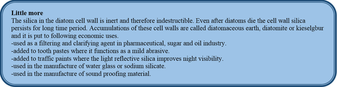
2.4.3 Divison - Euglenophyta (Euglenoids)
It is a group flagellated protistans that includes Euglena and euglena like (euglenoid) unicellular forms. Euglena is a taxonomic puzzle since it shows plant and animal characters. Absence of cell wall is an animal feature and presence of chloroplast a plant character of Euglena.
The cells are surrounded by a pellicle or periplast made up of proteins which form oblique lines of strip like myonemes. Pellicle is flexible so the cell can change its morphology.
Euglena has one flagellum per cell.
The cell can show holozoic nutrition. Cell of Euglena can ingest small sized food particles via cell mouth (cytostome) which leads to a cytopharynx (gullet). It also shows autotrophic nutrition by photosynthesis (myxotrophic nutrition).
Near the anterior end there is an eye spot or stigma which is a photoreceptor, it assists the cell to swim towards light and there are also contractile vacuoles for osmoregulation. There are many disc or ribbon shaped chloroplasts in a cell with photosynthetic pigments like chlorophyll a, b and carotenoids. Food is stored in the form of paramylum (a type of carbohydrate). The chloroplast have pyrenoids. The food matter is stored around the proteinaceous pyrenocrystal of the pyrenoid.
Cells are uninucleate and all eukaryotic organelles are present.
Euglenoids reproduce by longitudinal binary fission.
Sexual reproduction does not occur. Under unfavourable environmental conditions, like lack of water they form perennatory structures like the Cysts, when favourable conditions arrive the cyst produces the organism.
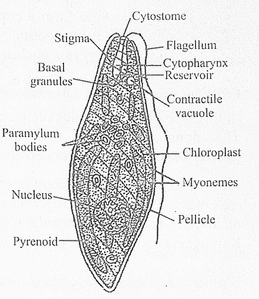
Fig.-2.18 Euglena
2.2.1 Slime moulds (protists which are consumers and decomposers)
They are found in terrestrial and aquatic habitats where they live on organic matter as a source of food. They can also ingest smaller organisms and behave as a consumer.
The organism is a slimy mass of protoplasm with animal and fungal characters. They are called mycetozoa or fungi like animals. They are also called fungal protists (True fungi belong to another kingdom called Mycota).
Slime moulds do not have cell wall, have amoeboid nature, holozoic or phagotrophic nutrition, spore wall does not have chitin and spores help in a sexual reproduction. Sexual reproduction is by fusion of flagellated gametes called swarm cells.
There are two types of slime moulds
a) Acellular slime moulds (Class - Myxomycetes)
b) Cellular slime moulds (Class - Acrasiomycetes)
(a) Acellular slime moulds
The organism has a plasmodium which is multinucleate protoplasm without walls. Common examples are Physarum and Stemonitis. The slimy plasmodium moves about in the substratum like an Amoeba feeding on organic debris or even bacteria.
When the environment becomes dry the plasmodium produces stalked sporangia. The diploid nuclei undergo meiosis in the sporangia where haploid spores are formed. Spores have galactose amine containing cell wall. The sporangium has protoplasmic threads called capillitia that helps in spore dispersal from the sporangium. The spores produce myxamoebae (amoeboid cells) or flagellated swarm cells.
The myxamoebae form a population of haploid cells. The haploid cells fuse to form diploid zygotes. The zygote germinates to form a plasmodium which becomes multinucleate (with diploid nuclei) by free nuclear mitotic divisions.
(b)Cellular slime molds (Acrasiomycetes)
In this type of slime moulds uninucleate amoeboid cells come together to form a psuedoplasmodiunt. The common example is Dictyostelium. The individual protoplasts do not fuse, therefore, it is a I psuedoplasmodium which is called slug form.
The psuedoplasmodium grows and forms sporangia. The sporangium produces spores with a wall of cellulose. The spores germinate to form myxamoebae, if the condition is dry myxamoebae form microcysts, in favourable condition the microcysts produce myxamoebae which produces macrocysts. Karyogamy and meiosis occur in macrocyst which produce haploid myxamoebae
Significance of pseudoplasmodium-Cellular slime mould demonstrates multicellularity of a primitive type where cells come together but maintain their uniqueness. Some cells form slugs and then sporangia. These organisms are highly evolved protists showing primitive fungal characters.
2.4.5 Division – Protozoa
Note: A detailed description of Protozoa is given in chapter 4.
2.5 KINGDOM MYCOTA (FUNGI)
The kingdom has more than one lakh species and all are heterotrophic, the nutrition is mostly saprophytic, parasitic and rarely symbiotic as in case of lichens and mycorrhizae, where fungal mycelium helps the root to absorb water and salts, and the roots provide organic food to the fungus.
Fungi can be described as achlorophyllous, heterotrophic, spore producing, chitinous cell wall containing non vascular eukaryotes which store reserve food as glycogen.
Sex organs when present are unicellular and zygotes undergo meiosis, therefore no embryos are produced.
They have developed a wide range of heterotrophic nutrition like saprophytic, parasitic and symbiotic (earlier explained)
Saprophytic nutrition: The fungi are decomposers or saprophytes that can breakdown a variety of organic macromolecules present is dead part of organisms and animal excreta. The extracellular enzymes are cellulases, proteases and lipases which breakdown cellulose, proteins and lipids respectively and from these macromolecules the simple building blocks produced, are easily absorbed by fungi to be assimilated for their growth.
Parasitic fungi obtain organic food from their hosts. The fungal plant parasites produce haustoria which penetrate the host cells to absorb organic food.
Fungal structure
The fungal body has thread like structures called hyphae (sing., hypha). Numerous hyphae are twined around one another to form many mycelia (sing; mycelium). Fungi like yeasts are unicellular, occasionally they from chains of cells by budding, these chains are psuedomycelia.
The hypha (are cylindrical) can be aseptate with many nuclei and all eukaryotic organelles are present (except plastids). The hypha shows localised apical growth. Such a aseptate and multinucleate structure is called a coenocyte. The hyphae could also be septate (with pores in the septum for cytoplasm to move between cells) with uninucleate or binucleate cells
When binucleate, the two nuclei in a cell look alike but they belong to + and - strain, hence it is called dikaryotic.
This is a secondary hypha produced by sexual fusion of only cytoplasm or plasmogamy of two primary + and - hyphae, nuclear fusion does not occur.
The hyphae are attached to the substratum rich in organic food by rhizoids. The hyphae can absorb food from the substratum.
All fungi pass through two phases or stages in their life cycle.
Vegetative or assimilative phase: During this phase they grow profusely in the food giving substratum which could be bread, cheese, wood. Coprophilous fungi grow well on cow dung, dead parts of plants like fallen leaves. They decompose leaves into mineral rich humus, dead bodies and excreta
Reproductive phase: In this phase, from the horizontal hyphae erect hyphae develop which produce reproductive structures. In unicellular yeasts, the entire cell become a reproductive structure, This is the hoiocarpic condition. In other fungi, reproductive structures develop from a part of the vegetative body and this condition is called eucarpic.
Reproduction in fungi occurs by vegetative means like fragmentation of hypha, asexual reproduction by motile spores (zoospores) or non - motile spores (aplanospores).
Sexual reproduction is by gametic fusion or hyphal fusion, gametes are produced in unicellular sex organs antheridia and oogonia, the male and female sex organs respectively. Gametic fusion results is the formation of diploid zygotes. The zygote divides meiotically. Embryo formation does not occur in fungi.
Cell and tissue level organisation
The cell wall is uniquely made up of chitin or fungal cellulose (in some primitive fungi like Pythim cellulose occurs in cell wall). The cell structure is typically eukaryotic with well formed nuclei ami chromosomes with histone proteins and DNA. The ribosomes in the cytoplasm are of 60S and 40S subunits which associate during protein synthesis to form 80S ribosomal complexes. Membrane bound organelles like mitochondria, lysosomes, dictyosmes of simple or unicistemal type is present. True vacuoles, endoplasmic reticulum are also present.
The mycelia often aggregate into tissue like structures like prosenchyma and psuedoparenchynta which are the two types of plectenchyma.
In prosenchyma many hyphae come close together and are parallely arranged.
In psuedoparenchyma the hyphae are closely packed. These tissues do not have cells interconnected with plasmodesmata like parenchyma tissues of vascular plants.
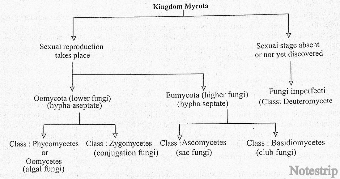
2.5.1 Class - Phycomycetes
They include primitive fungi, also called algal fungi, (Gr., phyco = algae; mycetes = fungi). The thallus (plant body) is made of up of cylindrical aseptate, branched, coenocytic hyphae with all eukaryotic organelles. Some oomycetes are unicellular. The cell wall has cellulose. The aquatic oomycetes are called water molds, some of them are saprophytes and some like the fish mold Saprolegnia may parasitise fish (it is a very common problem in all types of aquaria). The cottony mycelia of Saprolegnia covers the fins and body of fishes and kills the fishes. Some like Albugo Candida are parasites on angiosperm plants like mustard, causing white rust disease, the leaves develop white patches. Pythium debaryanum causes damping off disease in bean, mustard, and tomato plants the leaves are highly affected. The seedlings collapse to the soil, Phytophthora infestans causes late blight of potato plants and also tomato plants. Peronospora parasitica causes downy mildew disease in onion, pea and mustard plants. Plasmopara viticola causes downy mildew in grapes.
Asexual reproduction - This occurs by spores produced inside sporangia. Terrestrial species produce non motile aplanospores in aplanosporangia. The aquatic species produce motile or flagellate zoospores in zoosporangia. The zoospores can be uni or biflagellate. The biflagellate zoospore is a heterokont since one flagellum is of whiplash type and the other of the tinsel type with side branches.
Asexual reproduction also takes place by cells called oidia, these cells are produced when the fungi grow in a medium containing large quantities of sugars and acidic in nature. During oidia formation the hyphae becomes septate and produce many independent cells called oidia. The oidia detach and multiply by budding, when the buds fall on suitable substratum they develop into vegetative hyphae.
During unfavourable enviommental conditions numerous thick walled perennatory spore, called chlamydospores are produced by the hyphae. When favourable conditions return these spores develop into vegetative hyphae. They are called perennatory spores because they help the survival of organism during unfavourable enviommental conditions.
Sexual reproduction is by fusion of similar gametes (isogamy) or dissimilar gametes (anisogamy). The antheridia (male) and oogonia (female) are the unicellular sex organs. In isogamy the fusing gametes are identical to one another. In oogamy the male gamete is small and flagellate and female gamete is the large and immobile egg or ovum. The diploid zygote develops haploid zoomeiosporesby reduction division or meiosis (embryo is absent).
2.5.2 Class-Zygomycetes
They are also called conjugation fungi. Some of the examples are: Rhizopus and Mucor which cause spoilage of bread, pickles etc., The hyphae resemble those of the phycomycetes. Asexual reproduction is by spores produced in sporangia which develop on erect sporangiophores. Each sporangium has a central sterile zone called columella and around the columella is a fertile zone with numerous aplanospores or sporangiospores which are dispersed by wind. The hundreds of spores produced by a sporangium are liberated and they develop on the organic substrata, producing numerous hyphae rapidly. Sporangia and spores spoil food items.
Sexual reproduction : Rhizopus shows heterothallism where conjugation occurs between + and - strain hypha only. Heterothallism was discovered in 1904 by A.F. Blakeslee where conjugation occurs between + and - strain hyphae only, the two types of hyphae look similar but they are different genetic and physiological types. Multinucleate gametangia (coenogametangia) are produced on special hyphae called zygophores. The common wall between coenogametangia dissolves, the numerous + and - nuclei pair and fuse. The diploid nuclei undergo meiosis to form + and — haploid nuclei. The zygospore has many + and - nuclei. In some zygospores + nuclei degenerate and in some - nuclei degenerate.
The zygospore germinates during favorable environmental conditions to form a promycelium which terminates in a germsporangium. Different germ sporangia produce + and — strains pf germspores which produce + and - hyphae
2.5.3 Class - Ascomycetes
They are referred to as sac fungi because during sexual reproduction they produce sac like structures, called ascus (plu ; asci) in which generally eight ascopores developed by meiosis. Most of them are saprophytes, those which grow on cowdung and are called coprophilous. Some are parasites. The hypha is septate and the septa are perforated so that cytoplasm can pass from cell to cell. Some are unicellular like Saccharomyces (yeasts or sugar fungi).
The ascomycetes also produce unicellular sex organs, the female sex organs are called ascogonia (sing - ascogonium) and male sex organs are called antheridia (sing - antheridium). The ascomycetes includes fungi like Penicillium from which the antibiotic penicillin is obtained, the ergot fungus Claviceps purpurea produces fruiting bodies from which ergolin drug is obtained which is used to treat medical problems like migraine, palpitations of heart, menopausal disorders. The fungus Neurospora crassa has been used by biochemists and geneticists in research work. Ascomycetes includes morels and truffles whose fruiting bodies are edible. The ergot fungus attacks rye plant and consumption of ergot injected rye flour can kill humans by affecting the central nervous system causing death by convulsions. Many species of Aspergillus and Penicillium spoil food items, Aspergillus flavus infects red chillies and ground nuts and secrete aflatoxins which are harmful to humans as carcinogens.
2.5.4 Class - Basidiomycetes
The class includes fungi commonly called club fungi. They produce club shaped basidia (sing., basidium) and each basidium produces four sexual spores called basidiospores externally. The class includes saprophytes like mushrooms, bracket fungi, puffballs and parasites like rust and smut fungi.
The hypha is septate with a pore in the septum. The primary mycelia are produced by the germination of basidiospores. There are two strains (mating types) of basidiospores namely + and - strains (= heterothallism). The cells of the primary mycelia are uninucleate.
Asexual reproduction is absent and vegetative reproduction is by fragmentation of the thallus
Sexual reporduction- Sex organs are absent but there is a sexual mechanism of reproduction when + and - strain mycelial cells fuse to form a septate secondary mycelium of binucleate cells. Since each cell has a + and - nucleus the condition is called dikaryotic. During fusion of + and - cells there is only fusion of cytoplasm (plasmogamy). The secondary mycelia grow as saprophytes in the rich organic substrata like bark of trees and straw and produce fruiting bodies or basidiocarps. In the basidiocarps numerous club like structures called basidia develop. Inside the basidium, karyogamy takes place and the two (+ and -) nuclei fuse to form a diploid nucleus (diploid zygote) which undergoes reduction division or meiosis to form four haploid nuclei (two + and two - types). Each basidium produces four Anger like projections called sterigmata and each of them produces four basidiospores external to basidium. Each basidium produces two + strain or types of basidiospores and two - strain or type of basidiospores. The basidiospores are uninucleate cells which germinate to form the primary mycelia. (Fig. 2.28).
Mushrooms belonging to different genera like Agaricus, Pleurotes, Auricularia produce edible basidiocarps for human consumption (Amanita is a poisonous mushroom).
Edible mushrooms are being cultivated commercially. By taking directions from your teachers you can cultivate mushrooms on a small scale in your biology laboratory, it will be an interesting experience, if you follow the steps.
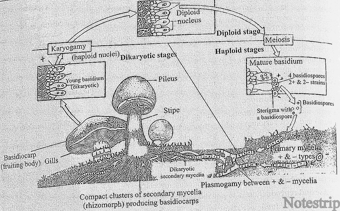
Fig.-2.19 Life cycle of a mushroom (agaricus)
Parasites like Puccinia graminis, causes black or red rust disease in wheat plants and Sphacelotheca sorghi (smut fungus) causes smut disease in sorghum plants. These diseases cause severe economic losses to the fanners.
2.5.5 Class - Deuteromycetes
This class of fungi are also called fungi imperfecti, because the sexual stage of reproduction or the I perfect stage is absent (or not yet discovered). This group is a taxonomic puzzle since whenever the I sexual stage is discovered in a fungus of this class, it is shifted to some other class of Mycota.
The hyphae are septate and uninucleate. The mode of nutrition is saprophytic or parasitic. 1 reproduction is asexual by means of conidia which infect the host plants.
Cercospora personata and Cercospora arachidicola attack leaves of ground nut plants causing I tikka or leaf spot disease.
Economic importance of fungi
(1) Useful fungi
(i) Fungi as food: Fungi like mushrooms are used as food. Yeasts also have great food value as I they are rich source of vitamin B. Dried yeasts contain about 50% protein.
(ii) Fungi in medicine: The first antibiotic penicillin, the wonder drug, was obtained from Penicillium notatum.
Some important medicines from fungi are as follows:
Penicillin Penicillium notatum, P. chrysogenum
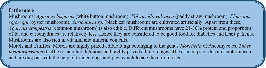
Ergotine : (Given after child birth to stop bleeding, to relieve migraine headaches and to treat hypertension.) Claviceps purpurea (Ergot).
Griseofulvin : (In skin diseases like ringworm) Pencillium griseofulvum
Fumigallin : Aspergillus fumigatus
Viridin : Gliocladium virens
Ramycin : Mucor ramannianus
Calvacin : (An anticancer substance) Clavatia sp.
Citrinine : P. citrinum
Patulin : Aspergillus clavatus
(iii) Fungi in industry:
Invertase: Saccharomyces, Altemaria
Zymase : Saccharomyces cerevisiae
Riboflavin (B12) | Saccharomyces cerevisiae
Vitamin (B12) Eremothecium ashbyii
Vitamin A Rhodotorula gracilis
2.5 KINGDOM - PLANTAE
The kingdom comprises chlorophyll containing photosynthetic eukaryotes with a cellulosic cell wall and a tissue level of organisation. A few plants are parasitic (since they lack chlorophyll)'. Some an I photosynthetic and carnivorous. Carnivorous plants like Nepenthes (pitcher plant) captures and digest insects to obtain nitrogenous compounds. Plantae or Metaphyta includes the higher algae (chlorophyte; 1 phaeophytes and ihodophytes), bryophytes, pteridophytes, gymnosperms and angiosperms.
There are two phases in the life cycles namely the diploid multicellular sporophyte and the 1 haploid multicellular gametophyte. These two phases alternate with one another in a phenomenon called alternation of generations. The duration of the two generations, their independent orjdependent nature varies among different groups. Details of these difference will be studied in chapter 3.
2.6 KINGDOM ANIMALIA
The kingdom includes heterotrophic eukaryotic organisms with cells without a cell wall and cells organised into tissues, organs and organs into different types of systems like digestive, nervous, I excretory, respiratory and locomotory. All animals depend upon plants directly or indirectly for food! The mode of nutrition is holozoic, they ingest solid food which is digested in the alimentary cattail. They have a programmed growth pattern and develop into adults. Reproduction is by sexual process 1 involving copulation of males and females followed by embryo development. Kingdom animalia is I divided into many phyla, (The details of the kingdom you will study in chapter 4).
2.8 PRIONS , VIROIDS, VIRUSES AND LICHENS
All these forms are not included in any of the five kingdoms since they have highly unique combinations of characters.
2.8.1 Prions
They are infections particles made up of proteins (They have no nucleic acids) However the proteins of prions (pronounced as preeons) infect animals like cattle and humans.
All the presently known prion caused diseases are untreatable and fatal and these diseases are called transmissible spongiform encephalopathies (TSEs).
The disease scrapie in sheep (sheep scrape their bodies on rocks causing wool loss), mad cow disease or bovine spongiform encephalopathy in cattle (BSE) and Creutzfeldt Jacob disease and Kuril disease in humans are caused by prions.
2.8.2 Viroids
The viroids are naked circular single stranded RNA (ssRNA) molecules without any protein coat. Diener discovered them in 1967.
They are simpler than viruses. The RNA of the viroid has 250 to 370 nucleotides, the RNA can reproduce only in living host cells.
2.8.3 Viruses
Viruses are unique infectious agents, this is the reason why they have not been taken into Whittaker’s I system of classification in any kingdom.
Louis Pasteur coined the term virus (L. virus = poison).
Viruses are ultramicroscopic, noncellular, nuceloprotein particles with only one type of nucleic I acid (Deoxyribonucleic Acid, DNA or Ribonucleic acid, RNA) and they are obligate, intracellular I parasites in living host cells.
2.8.4 Lichens
These organisms have two components namely algal component (phycobiont) and fungal component (mycobiont). The mycobiont could be the ascomycete (ascolichen) or basidiomycete (basidiolicben) fungus, where as the phycobiont could be cyanobacteria (blue green alge) like Anabenciy Nostoc or Gloeocapsa, or green algae (Chlorophyceae), some of which are Protococcus and Trentepholia. The major part of the lichen body is composed of fungal mycelia.
The relationship between the two components is mutually beneficial (mutualism) and is popularly known as symbiosis. The fungal mycelium fixes the lichen thallus to the substratum, it protects the algal cells from dehydration and provides algal cells with water and mineral salts. The algal cell by photosynthesis provides organic food to the fungi and if the algae is a nitrogen fixing cyanobacterium the fungal cells also get useful nitrates.
Lichens can tolerate extremely hot and dry conditions. They can grow on wide range of habitats- the surface of a bare rock (lithophytes) and on tree barks (epiphytes). They occur even in cold icy tundra regions. As a taxonomic entity lichens are a large group of 400 genera and 15,000 species. Lichens grow very slowly and live long. Some lichens of the arctic regions are 4,500 years old.
Lichens growing on rocks are called saxlcoles, those growing on tree barks are called corticoles and lichens growing on soil are called terricoles.
Griseofulvin : (In skin diseases like ringworm) Pencillium griseofulvum
Fumigallin : Aspergillus fumigatus
Viridin : Gliocladium virens
Ramycin : Mucor ramannianus
Calvacin : (An anticancer substance) Clavatia sp.
Citrinine : P. citrinum
Patulin : Aspergillus clavatus
(iii) Fungi in industry:
- Bakervs yeast (Saccharomyces cerevisiae) is used in baking industry.
- Enzyme production from fungi
Invertase: Saccharomyces, Altemaria
Zymase : Saccharomyces cerevisiae
- Vitamins from fungi:
Riboflavin (B12) | Saccharomyces cerevisiae
Vitamin (B12) Eremothecium ashbyii
Vitamin A Rhodotorula gracilis
2.5 KINGDOM - PLANTAE
The kingdom comprises chlorophyll containing photosynthetic eukaryotes with a cellulosic cell wall and a tissue level of organisation. A few plants are parasitic (since they lack chlorophyll)'. Some an I photosynthetic and carnivorous. Carnivorous plants like Nepenthes (pitcher plant) captures and digest insects to obtain nitrogenous compounds. Plantae or Metaphyta includes the higher algae (chlorophyte; 1 phaeophytes and ihodophytes), bryophytes, pteridophytes, gymnosperms and angiosperms.
There are two phases in the life cycles namely the diploid multicellular sporophyte and the 1 haploid multicellular gametophyte. These two phases alternate with one another in a phenomenon called alternation of generations. The duration of the two generations, their independent orjdependent nature varies among different groups. Details of these difference will be studied in chapter 3.
2.6 KINGDOM ANIMALIA
The kingdom includes heterotrophic eukaryotic organisms with cells without a cell wall and cells organised into tissues, organs and organs into different types of systems like digestive, nervous, I excretory, respiratory and locomotory. All animals depend upon plants directly or indirectly for food! The mode of nutrition is holozoic, they ingest solid food which is digested in the alimentary cattail. They have a programmed growth pattern and develop into adults. Reproduction is by sexual process 1 involving copulation of males and females followed by embryo development. Kingdom animalia is I divided into many phyla, (The details of the kingdom you will study in chapter 4).
2.8 PRIONS , VIROIDS, VIRUSES AND LICHENS
All these forms are not included in any of the five kingdoms since they have highly unique combinations of characters.
2.8.1 Prions
They are infections particles made up of proteins (They have no nucleic acids) However the proteins of prions (pronounced as preeons) infect animals like cattle and humans.
All the presently known prion caused diseases are untreatable and fatal and these diseases are called transmissible spongiform encephalopathies (TSEs).
The disease scrapie in sheep (sheep scrape their bodies on rocks causing wool loss), mad cow disease or bovine spongiform encephalopathy in cattle (BSE) and Creutzfeldt Jacob disease and Kuril disease in humans are caused by prions.
- Stanley B Prusiner discovered prions in 1983 (Awarded Nobel prize in 1997)
- Prions do not cause plant diseases. They cause fatal disease in cattle, sheep and humans. Symptoms appear years after infection. The brain is damaged by destruction of neurons and small cavities appear in brain, this makes the brain spongy (spongiform).
- Prions are unaffected by chemicals, heat and radiation.
- The aggregated prion protein in brain causes neurological damage.
2.8.2 Viroids
The viroids are naked circular single stranded RNA (ssRNA) molecules without any protein coat. Diener discovered them in 1967.
They are simpler than viruses. The RNA of the viroid has 250 to 370 nucleotides, the RNA can reproduce only in living host cells.
2.8.3 Viruses
Viruses are unique infectious agents, this is the reason why they have not been taken into Whittaker’s I system of classification in any kingdom.
Louis Pasteur coined the term virus (L. virus = poison).
Viruses are ultramicroscopic, noncellular, nuceloprotein particles with only one type of nucleic I acid (Deoxyribonucleic Acid, DNA or Ribonucleic acid, RNA) and they are obligate, intracellular I parasites in living host cells.
2.8.4 Lichens
These organisms have two components namely algal component (phycobiont) and fungal component (mycobiont). The mycobiont could be the ascomycete (ascolichen) or basidiomycete (basidiolicben) fungus, where as the phycobiont could be cyanobacteria (blue green alge) like Anabenciy Nostoc or Gloeocapsa, or green algae (Chlorophyceae), some of which are Protococcus and Trentepholia. The major part of the lichen body is composed of fungal mycelia.
The relationship between the two components is mutually beneficial (mutualism) and is popularly known as symbiosis. The fungal mycelium fixes the lichen thallus to the substratum, it protects the algal cells from dehydration and provides algal cells with water and mineral salts. The algal cell by photosynthesis provides organic food to the fungi and if the algae is a nitrogen fixing cyanobacterium the fungal cells also get useful nitrates.
Lichens can tolerate extremely hot and dry conditions. They can grow on wide range of habitats- the surface of a bare rock (lithophytes) and on tree barks (epiphytes). They occur even in cold icy tundra regions. As a taxonomic entity lichens are a large group of 400 genera and 15,000 species. Lichens grow very slowly and live long. Some lichens of the arctic regions are 4,500 years old.
Lichens growing on rocks are called saxlcoles, those growing on tree barks are called corticoles and lichens growing on soil are called terricoles.
